
|
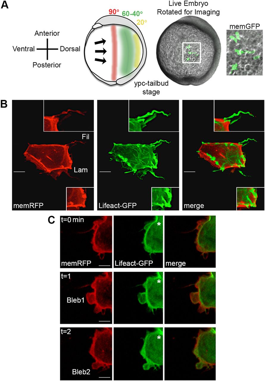
Fig. 1
Time-lapse imaging of membrane protrusions. (A) Schematic (left) and live wild-type zebrafish embryo (right) highlighting the 40°-60° lateral region that was analyzed. Lateral ectodermal cells with mosaic memGFP expression from an embryo used for confocal imaging at 60° from dorsal (white boxed area, enlarged far right). ypc, yolk-plug closure. (B) Wild-type ectodermal cell expressing memRFP and Lifeact-GFP with filopodia (Fil) and large lamellipodia-like (Lam) membrane protrusions. Insets show enlarged images of protrusion classifications. (C) Wild-type ectodermal cell undergoing division. Asterisks indicate forming cleavage furrow. Two blebs are observed during the three time points. Scale bars: 5 µm.