
Fig. 2
- ID
- ZDB-IMAGE-181127-14
- Publication
- Sun et al., 2018 - Mga Modulates Bmpr1a Activity by Antagonizing Bs69 in Zebrafish
- All Figures
- Figures for Sun et al., 2018
|
Fig. 2
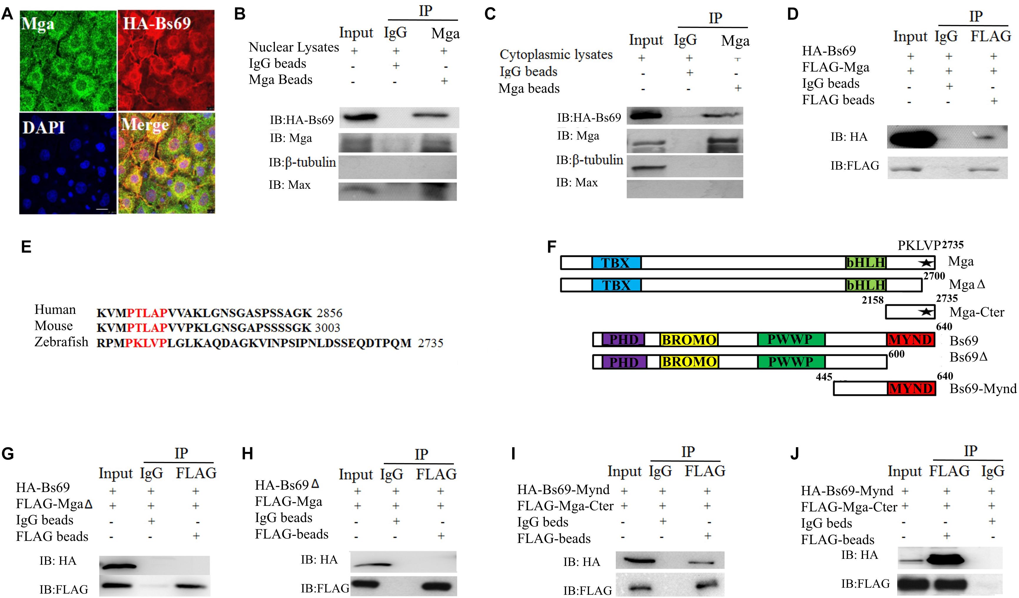
Mga interacts with Bs69. (A) Confocal images of Mga and HA-Bs69 proteins in 7 hpf embryos. Scale bar 10 μm. (B) Co-immunoprecipitation of Mga and HA-Bs69 in nuclear lysates from 8 hpf embryos. (C) Co-immunoprecipitation of Mga and HA-Bs69 in cytoplasmic lysates from 8 hpf embryos. (D) Co-immunoprecipitation of HA-Bs69 and FLAG-Mga in 293T cells. (E) Identification of a conserved PXLXP motif in human, mouse and zebrafish MGAs at the end of the C-terminal region. The PXLXP conserved motif were highlighted in red. (F) Schematic representation of the full-length and truncated Mga and Bs69 constructs as indicated. (G) FLAG-MgaΔ did not immunoprecipitate with HA-Bs69 in 293T cells. (H) FLAG-Mga did not immunoprecipitate with HA-Bs69Δ in 293T cells. (I) FLAG-Mga-Cter immunoprecipitated with HA-Bs69-Mynd in 293T cells. (J) Co-immunoprecipitation assay of the in vitro translated FLAG-Mga-Cter and HA-Bs69-Mynd.