
Fig. S6
- ID
- ZDB-IMAGE-181120-7
- Publication
- Earley et al., 2018 - Critical Role for a Subset of Intestinal Macrophages in Shaping Gut Microbiota in Adult Zebrafish
- All Figures
- Figures for Earley et al., 2018
|
Fig. S6
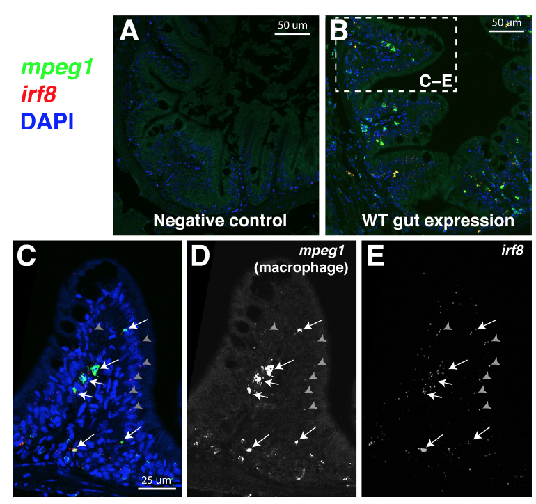
RNAscope in situ hybridization reveals restricted irf8 expression in intestinal macrophages in wild type adult zebrafish, Related to Figure 6.
Representative images of 5 μm paraffin transverse sections of the adult zebrafish through the intestinal region processed for RNAscope in situ hybridization. A Negative control multiplex RNAscope staining. B Merge fluorescent image showing a cross section of the adult intestine stained for mpeg1 (FITC, green) and irf8 (Cy3, red) gene-specific RNAscope probes with DAPI (blue) as a counterstain to visualize all cells. C High magnification of dotted box region in B shows expression of irf8 in the same cells also expressing the macrophage marker mpeg1 (arrows) in an intestinal villus fold. Arrows point to macrophages positive for both probes (mpeg1+ and irf8+). Gray arrowheads show representative punctate signals also found positive for both probes (mpeg1+ and irf8+). D Grayscale image of mpeg1 (FITC) channel alone. E Grayscale image of irf8 channel (Cy3) alone.