
Fig. S1
- ID
- ZDB-IMAGE-181109-7
- Publication
- Romero et al., 2018 - Damage-induced reactive oxygen species enable zebrafish tail regeneration by repositioning of Hedgehog expressing cells
- All Figures
- Figures for Romero et al., 2018
|
Fig. S1
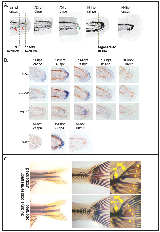
Larval tail regeneration. (A) When cut in the pigment gap (zone between the two red arrows), the notochord bead (the green arrow) is extruded within 3hpe and the tail regenerates within 4-5 days. The position of fin fold excision is shown for comparison. (B) Different stages of regeneration are marked by the expression of dlx5a, raldh2, myod and msxc (all panels 10/10 except dlx5a/24hpe 8/10, raldh2/24hpe 8/10, myod/48hpe 8/10, number of experiments = 3). Uncut fish are shown to the right for comparison. (C) Live images of the tail after metamorphosis is complete (left panels) and Von Kossa stain showing the failure of the notochord to extend dorsally (right panels). In unoperated fish the uroneural and hypurals 3-5 form around and are attached to the notochord which grows dorsally as the adult tail forms. After excision these bones all form on the urostyle suggesting that the dorsal flexure of the notochord has not occurred (both operated and unoperated are 5/5, number of experiments = 2). Abbreviations: urostyle (ust), uroneural (uro) and hypural 1-5 (1-5).