Fig. 4
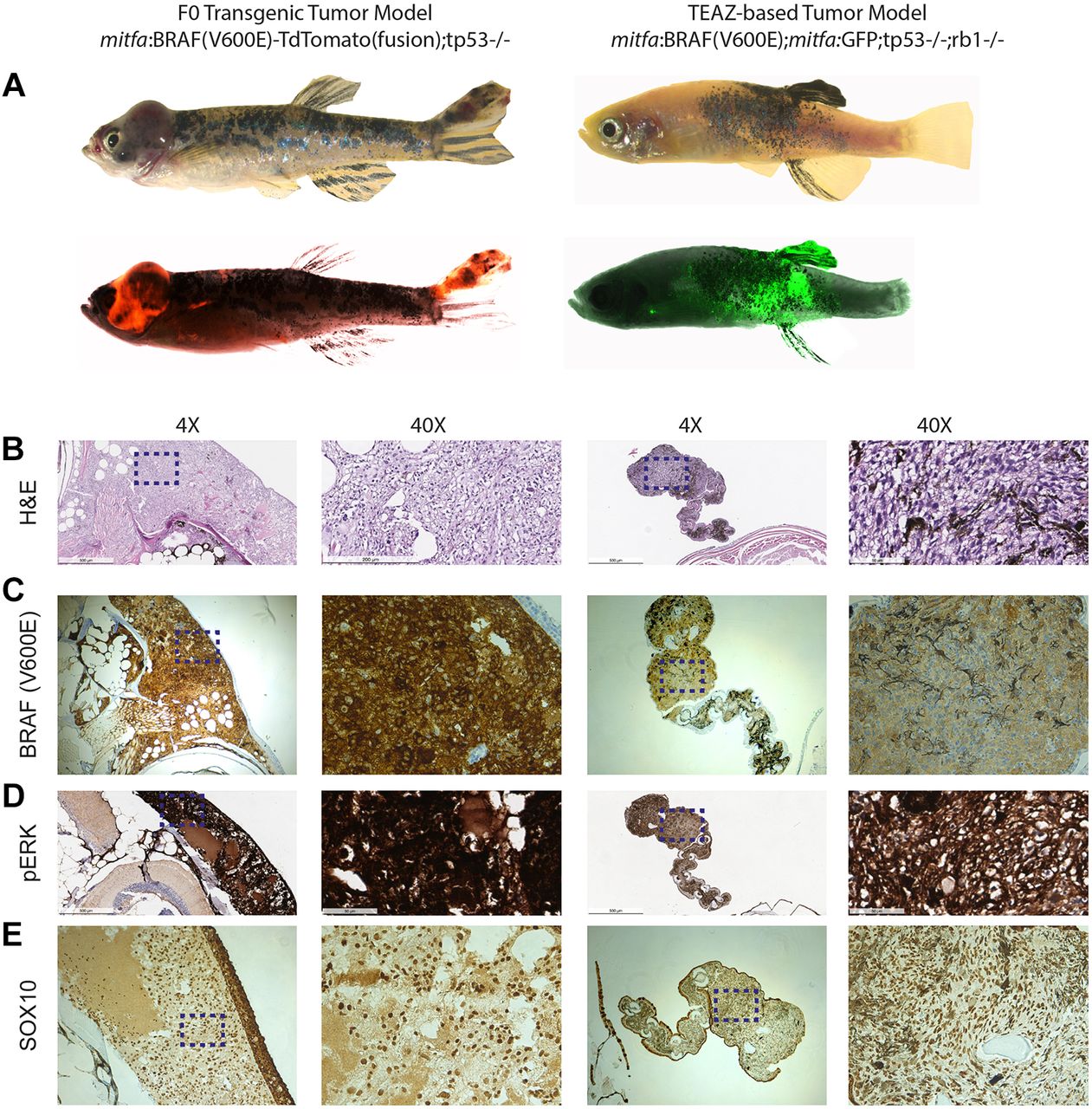
Histological comparison of the embryo injection melanoma model and TEAZ melanoma model. (A) The left images show a melanoma created by injection of an mitfa:BRAFV600E-tdTomato (fusion) transgene into a tp53−/− background (n=1). Right images show a TEAZ-based melanoma created by electroporation of miniCoopR:GFP plus ubb:Cas9 plus zfU6:sgRNA against Rb1 (n=1) (example shown is fish at 16 weeks also shown in Fig. 2A). (B) H&E staining of both tumors shows similar histology, although with increased melanin pigmentation in the TEAZ tumor (also shown in Fig. 3A). (C,D) Antibody staining against BRAFV600E shows that both tumors are widely BRAFV600E positive, which correlates with high levels of phospho-ERK staining. (E) Reflecting the neural crest origin of melanocytes, both tumors show strong nuclear expression of SOX10. Images are visualized at 4× and 40×. Scale bars: 500 μm (4×) and 50 μm (40×). Dashed line boxes indicate the area enlarged at 40×.

