Image
Figure Caption
Fig. 3
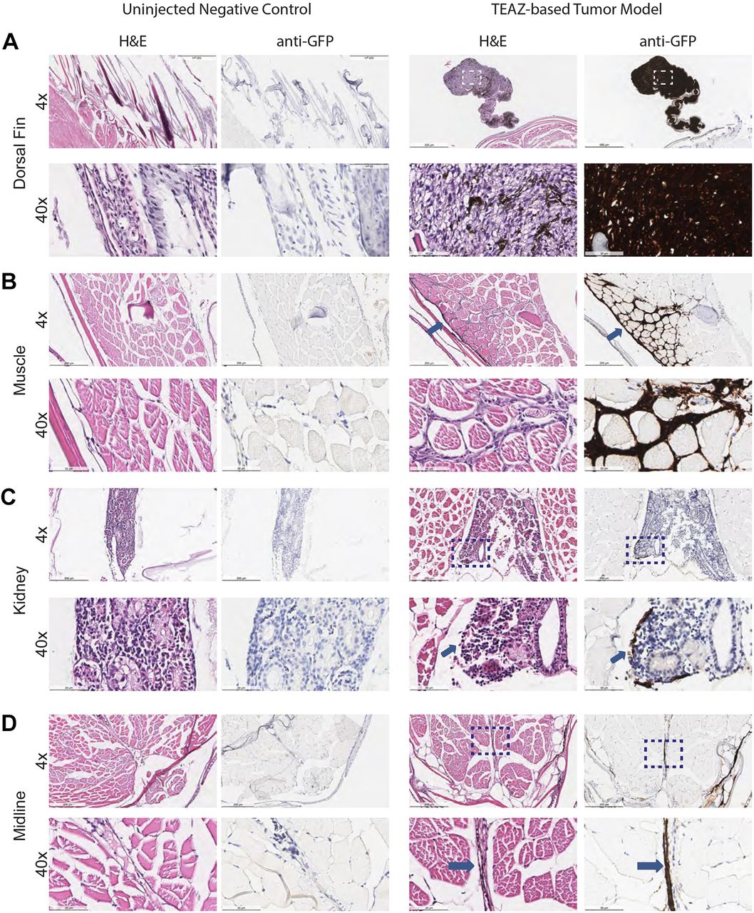
Melanoma model using TEAZ show evidence of rapid progression. (A) Pathology of tumor-bearing zebrafish (n=1) (along with control zebrafish, n=2) at 16 weeks postelectroporation, stained with H&E or anti-GFP immunohistochemistry, demonstrates a large primary tumor that is uniformly GFP+. (B-D) Histology reveals evidence of extensive invasion into the muscle (arrows) (B) along with micrometastatic sites within the kidney (C) or along blood vessels (arrows) (D). Images are visualized at 4× and 40×. Scale bars: 500 μm (4×) and 50 μm (40×). Dashed line boxes indicate the area enlarged at 40×.
Acknowledgments
This image is the copyrighted work of the attributed author or publisher, and
ZFIN has permission only to display this image to its users.
Additional permissions should be obtained from the applicable author or publisher of the image.
Full text @ Dis. Model. Mech.

