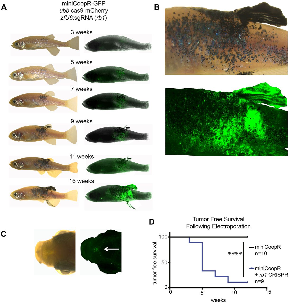
Fig. 2
Generation of a novel melanoma model with TEAZ. (A) mitfa:BRAFV600E;tp53−/−;mitfa−/− zebrafish (triple strain) were electroporated with the miniCoopR:GFP plasmid that both rescues melanocytes and expresses GFP under the mitfa promoter, with (n=10) or without (n=9) two additional plasmids to genetically knockout rb1 (ubb:Cas9 and zfU6:sgRNA against rb1). The electroporated zebrafish were then imaged over time by both fluorescence and brightfield to monitor tumor development. Overall, 17/20 electroporated zebrafish had GFP+ cells. Tumor development in a representative zebrafish from the melanoma model including rb1 knockout is shown. (B) Higher-magnification view of the tumor-bearing animal shown in A at 16 weeks postelectroporation. (C) At 9 weeks postelectroporation, 4/8 zebrafish had evidence of GFP+ distant micrometastases in the head. (D) The loss of rb1 is essential for tumor initiation as visualized by the Kaplan–Meier curve comparing zebrafish electroporated with miniCoopR:GFP with or without rb1 sgRNA. Log-rank (Mantel–Cox) test was used for statistical analysis (****P<0.0001).

