
|
Fig. 4
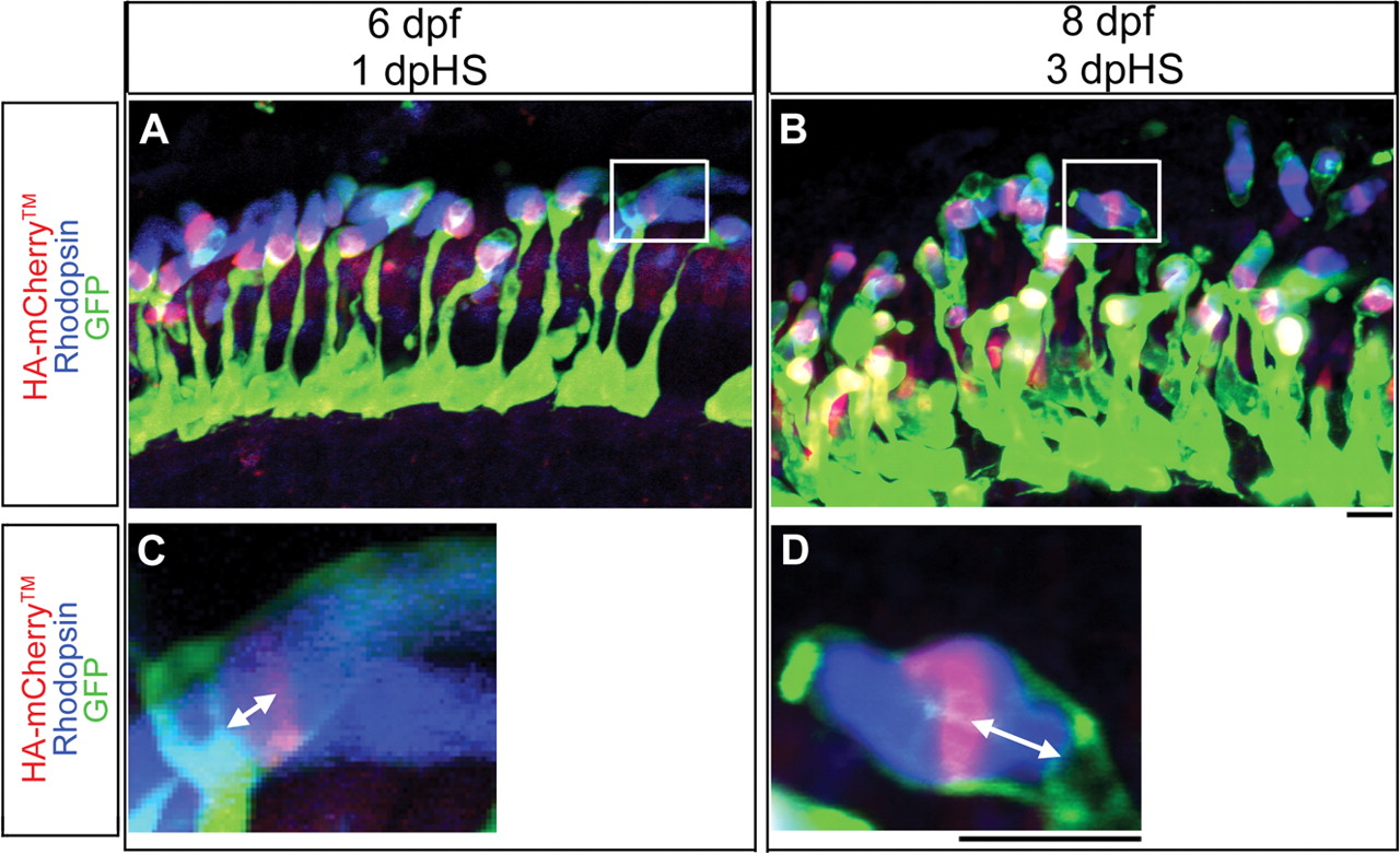
Expression of HA-mCherryTM in photoreceptors after heat-shock at 5 dpf.
Confocal z-projections of Tg(hsp70:HA-mCherryTM) photoreceptor layers labeled with anti-HA antibody (red), anti-Rhodopsin antibody (blue) and with GFP-expressing rods (green). (A) At 6 dpf, 1 dpHS, HA-mCherryTM in rod outer segments is seen as a stripe, an oval or a circle, depending on the orientation of the outer segment to the plane of section. (C) A magnification of the boxed area in (A) shows the displacement of the HA-mCherryTM stripe from the base of the outer segment in 2-dimensions. (B) At 8 dpf, 3 dpHS, HA-mCherryTM in rod outer segments is seen as a stripe, an oval or a circle, depending on the orientation of the outer segment to the plane of section and HA-mCherryTM has moved distally compared to that seen at 1 dpHS. (D) A magnification of the boxed area in (B) shows the displacement of the HA-mCherryTM stripe from the base of the outer segment in 2-dimensions. Scale bars: 5 µm, A, B; 5 µm, C, D.