
Image
|
Figure Caption
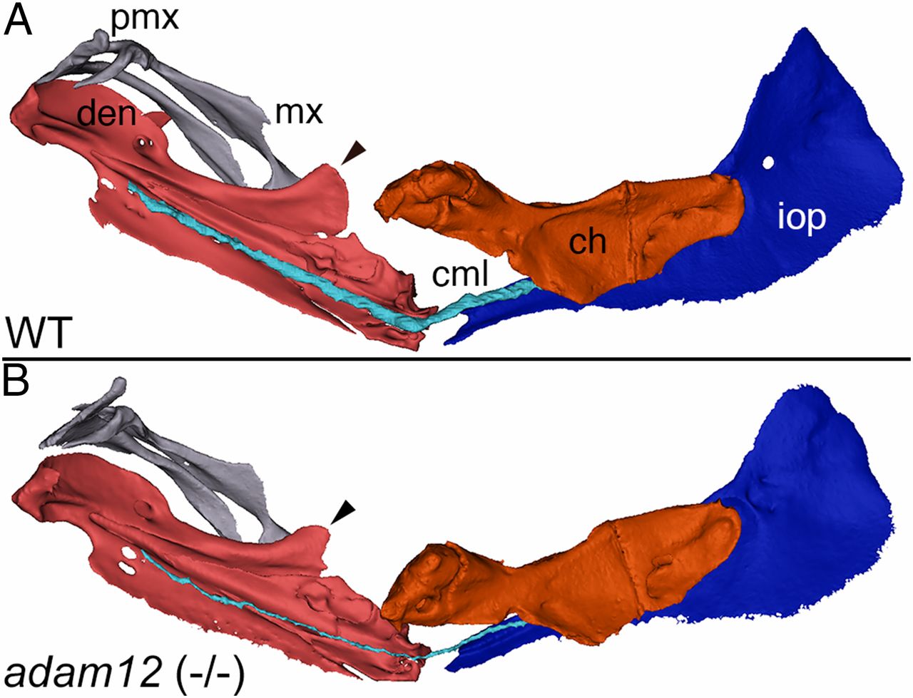
Fig. 6
µCT data demonstrate reduced ligament volume in adam12 zebrafish mutants. (A) Reconstructed 3D model in a wild-type zebrafish illustrating position of the cerato-mandibular ligament (cml) relative to mandible (den) and ceratohyal (ch). Maxilla (mx), premaxilla (pmx), and interopercle (iop) are also shown for perspective. (B) Reconstructed 3D model of the same structures in an adam12 mutant zebrafish. Note the marked reduction in the size of the cml (blue) and the coronoid process of the mandible (arrowheads).
Acknowledgments
This image is the copyrighted work of the attributed author or publisher, and
ZFIN has permission only to display this image to its users.
Additional permissions should be obtained from the applicable author or publisher of the image.
Full text @ Proc. Natl. Acad. Sci. USA