
|
Fig. 5-S1
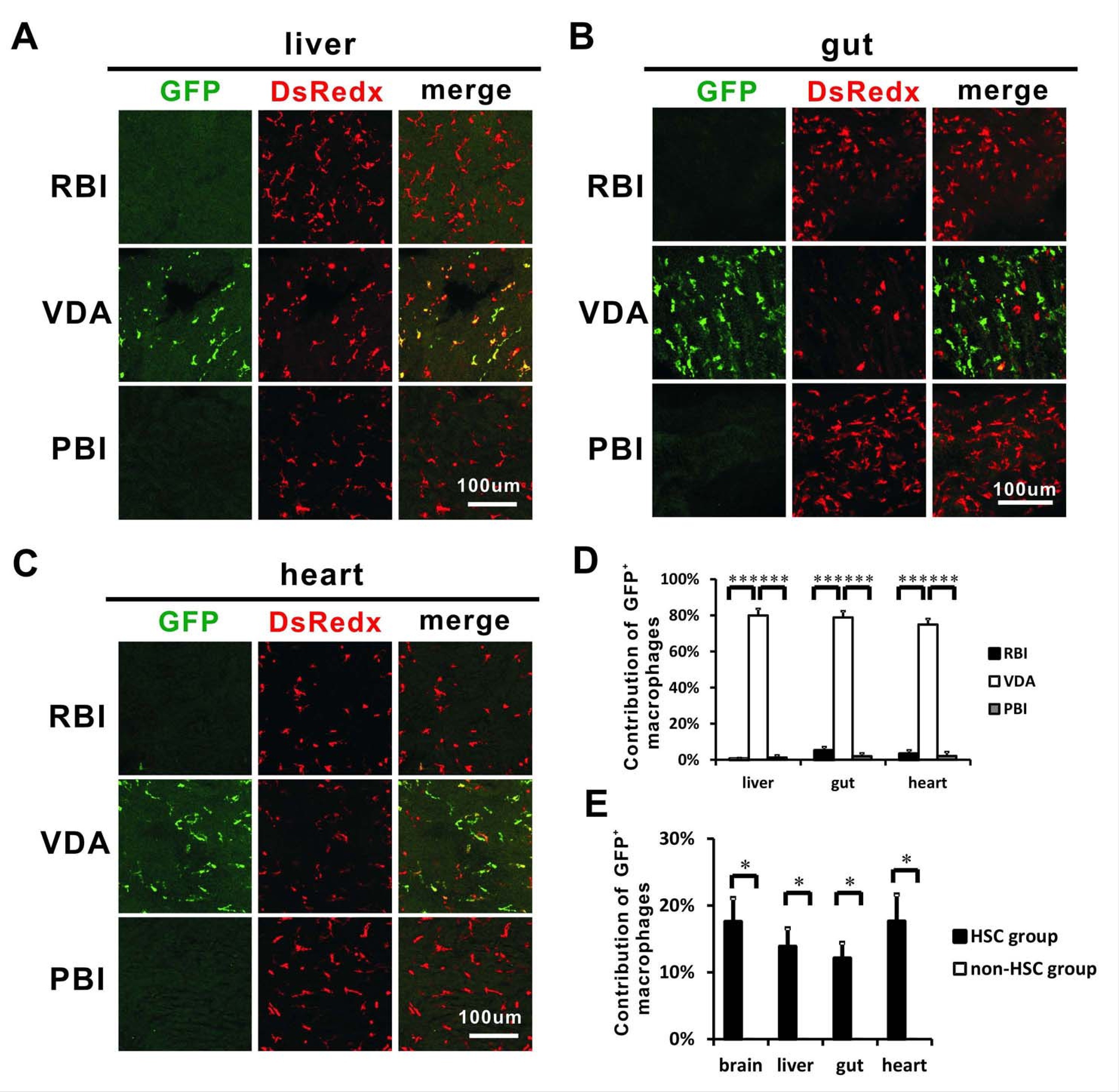
Other tissue-resident macrophages also correlate with HSCs.
(A) Anti-GFP staining indicates that GFP+ resident macrophages are mainly detected in the VDA-labelled fish, but not the RBI- and PBI-labelled fish in adult liver. (B) Anti-GFP staining indicates that GFP+ resident macrophages are mainly detected in the VDA-labelled fish, but not the RBI- and PBI-labelled fish in adult gut. (C) Anti-GFP staining indicates that GFP+ resident macrophages are mainly detected in the VDA-labelled fish, but not the RBI- and PBI-labelled fish in the adult heart. (D) Quantification of the percentage of GFP+ resident macrophages derived from the RBI, VDA, and PBI in adult liver, gut, and heart. n = 5 for each sample analyzed. Error bars represent mean SEM. ***p<0.001. (E) Quantification of the relative contribution of GFP+ cells in the adult liver, gut and heart of HSC (n = 4) and non-HSC (n = 3) groups. Error bars represent mean SEM. *p<0.05.