
|
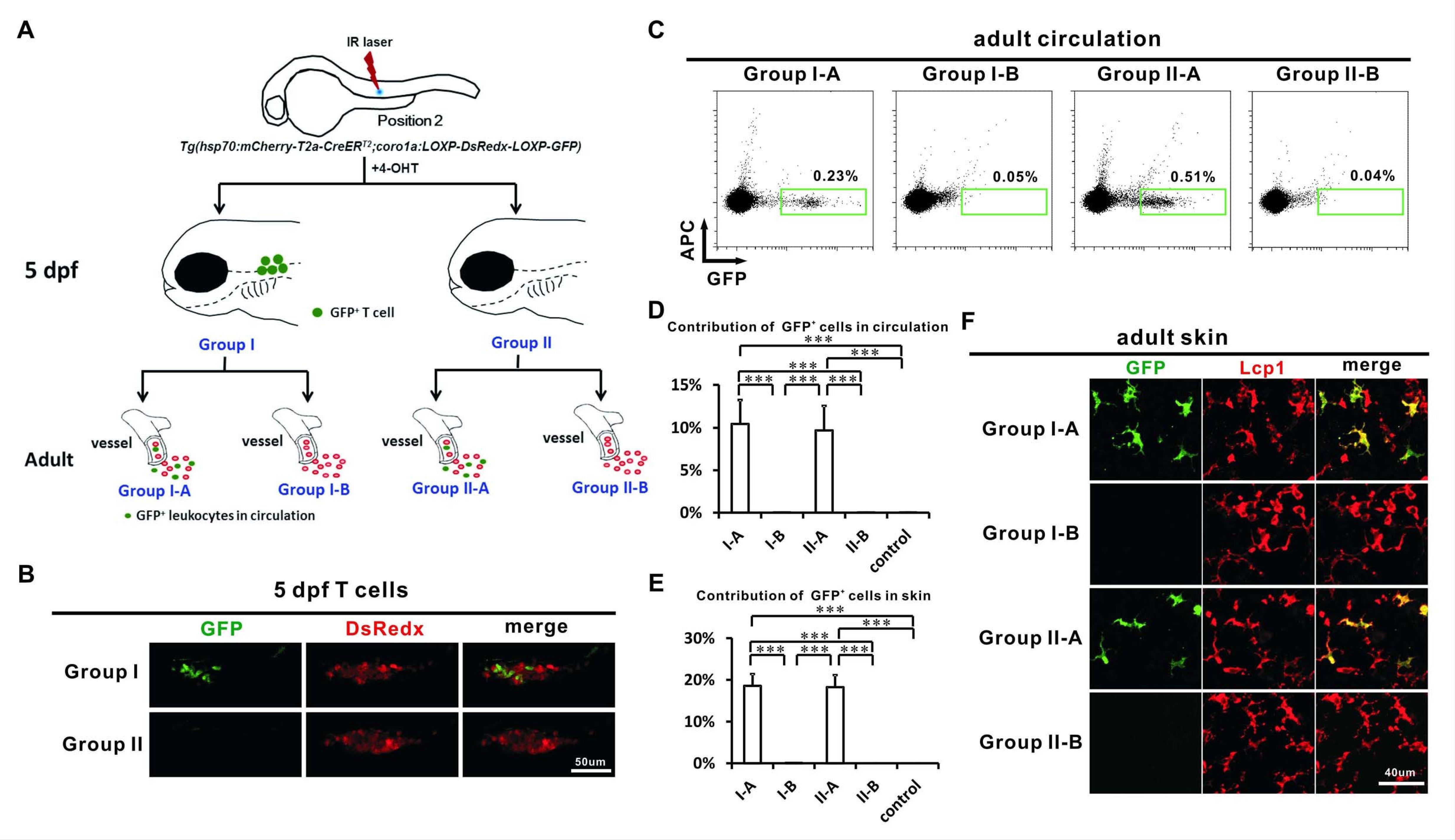
Fig. 4
Adult LCs correlate with HSCs but not non-HSC progenitors.
(A) A schematic view of the experimental design for laser labeling of Tg(hsp70:mCherry-T2a-CreERT2;coro1a:loxP-DsRedx-loxP-GFP) embryos. A single IR laser hit was performed at Position 2 of VDA at about 24 hpf. After 4-OHT treatment, the heat-shocked embryos were raised to 5 dpf and the embryos were then separated into two groups according the GFP contribution to thymocytes. Group I embryos contain abundant GFP+ thymocytes, whereas Group II embryos hardly have GFP+ thymocytes. These two groups of embryos were raised to adult (over 3 months) and were subdivided into Group I-A, I-B, II-A and II-B according to the GFP contribution in circulation. (B) Confocal images show that Group I embryos, but not Group II embryos, contain abundant GFP+ thymocytes. (C) Flow cytometry shows that the circulation of Group I-A and II-A, but not I-B and II-B contain abundant GFP+ cells. (D) Quantification of the contribution of GFP+ cells in all fluorescent positive leukocytes in the circulation of Group I-A, I-B, II-A, II-B, and non-heat-shocked control group (n = 6 for each group). Error bars represent mean SEM. ***p<0.001. (E) Quantification of the contribution of GFP+ cells in all Lcp1 positive leukocytes in the skin of adult Group I-A, I-B, II-A, II-B, and non-heat-shocked control group (n = 5 for each group). Error bars represent mean SEM. ***p<0.001. (F) Confocal images show that GFP+ cells are mainly found on the skin of Group I-A and II-A, but not I-B and II-B. a significant portion of GFP+ cells showed ramified LC-like morphology, suggesting that they are LCs presumably.