
|
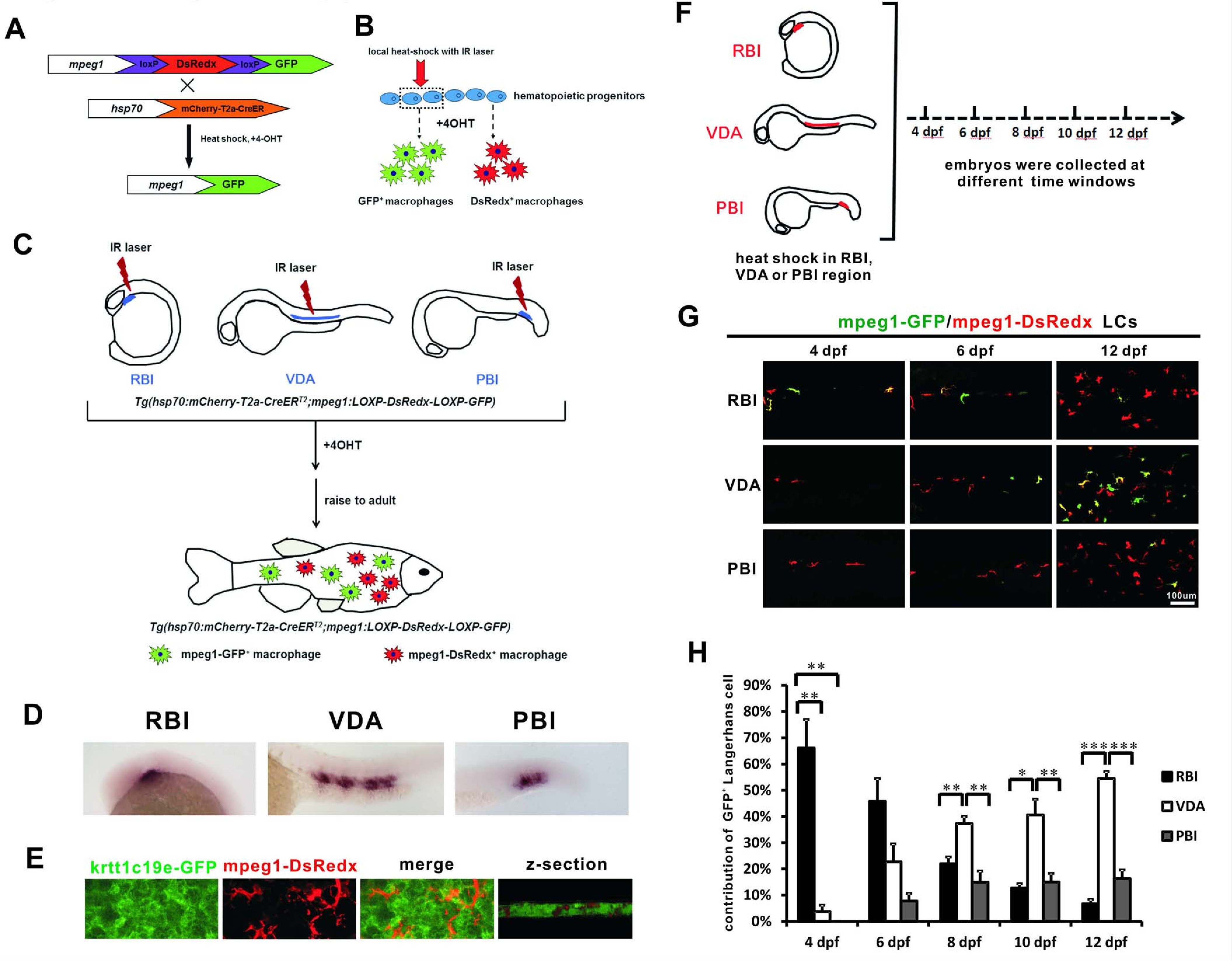
Fig. 1-S1
Experimental design of the IR-LEGO-CreER-loxP cell labelling system for LC fate mapping.
(A) The Tg(mpeg1:loxP-DsRedx-loxP-GFP) fish was crossed with the heat shock-inducible Tg(hsp70:mCherry-T2a-CreERT2) line. The resulted double transgenic embryos were applied to infrared heats-hock. Upon 4-OHT treatment, CreER mediated loxP recombination occurs. (B) Local heat-shock was introduced with IR laser to hematopoietic progenitors and 4-OHT was applied to induce loxP recombination. After cell differentiation, only the macrophages derived from the heat-shocked progenitors would express GFP, whereas the other macrophages remain DsRedx expression. (C) In the double transgenic embryos, IR laser was introduced to RBI, VDA or PBI, the three regions produce primitive myeloid cells, definitive hematopoiesis or intermediate EMPs respectively. After 4-OHT treatment, the embryos were raised to adult and the contribution of GFP+ and DsRedx+ LCs were examined. (D) Local heat-shock successfully induced CreER expression in RBI, VDA, and PBI. (E) mpeg1-DsRedx+ macrophages (red signals) locate within the krtt1c19e-GFP+ (green signals) basal epidermal layer as shown by the 3D view of the z-section. (F) A timeline shows that the fish heat-shocked in RBI, VDA and PBI regions were examined from 4 dpf to 12 dpf at 2 day intervals. (G) Imaging of the skin at 4 dpf, 6 dpf, and 12 dpf shows that VDA-derived GFP+ LCs gradually become the predominant population. The mpeg1-GFP and mpeg1-DsRedx signals were merged together, therefore both green and yellow LCs represent GFP+ cells. (H) Quantification of the percentage of RBI, VDA, and PBI derived LCs from 4 dpf to 12 dpf at 2 day intervals. n = 6, 5, and 5 for RBI, VDA, and PBI, respectively, (n = 4 for VDA at 10 dpf and 12 dpf). Error bars represent mean SEM. *p<0.05; **p<0.01; ***p<0.001.