
|
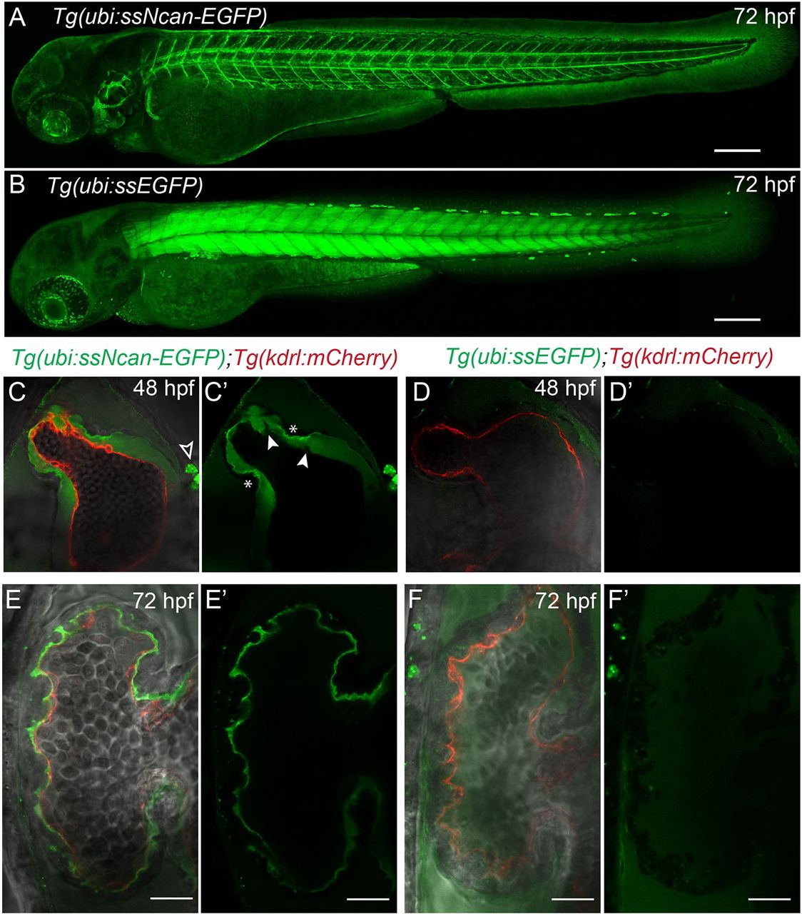
Fig. 3
A transgenic biosensor for the cardiac jelly. (A) Lateral view of the Tg(ubi:ssNcan-EGFP) line: a biosensor for hyaluronic acid – a major constituent of the cardiac jelly. The sensor expresses a secreted fusion protein of the HA-binding domain of Neurocan fused to GFP, driven by the ubi promoter. The biosensor localises to regions of HA, including the cardiac jelly, the otic vesicle and around the vasculature. (B) The Tg(ubi:ssEGFP) control was also generated and shows different EGFP localisation, predominantly to the skeletal muscle. Scale bars: 100 µm. (C,C′) Frontal view images of 48 hpf Tg(ubi:ssNcan-EGFP)/Tg(kdrl:mCherry) hearts anaesthetised and live imaged showing the cardiac jelly (green) and endocardial layer (red). ssNcan-EGFP localises to mucous cells (black arrowhead) and is concentrated at the AVC (asterisks). Regions devoid of ssNcan-EGFP in the cardiac jelly due to protruding endocardial cells can also be observed (white arrowheads). (D,D′) Live images of 48 hpf Tg(ubi:ssEGFP)/Tg(kdrl:mCherry) hearts showing near undetectable amounts of ssEGFP localisation (for full z-stacks of C,D, see Movie 2). (E,E′) Live images of 72 hpf Tg(ubi:ssNcan-EGFP)/Tg(kdrl:mCherry) ventricles showing condensed jelly and sites of trabeculae formation. (F,F′) Live images of 72 hpf Tg(ubi:ssEGFP)/Tg(kdrl:mCherry) hearts showing ssEGFP localisation in the pericardial space. Scale bar: 20 µm.