
Fig. 1
- ID
- ZDB-IMAGE-181018-6
- Publication
- Du et al., 2018 - A transgenic zebrafish model for in vivo long-term imaging of retinotectal synaptogenesis
- All Figures
- Figures for Du et al., 2018
|
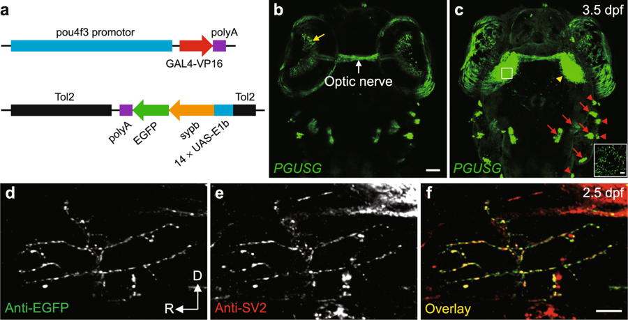
Fig. 1
Visualization of RGC pre-synaptic terminals in double transgenic zebrafish PGUSG. (a) Schematic of the DNA constructs used for generating the GAL4 line Tg(pou4f3:GAL4-VP16) (PG) and the UAS line Tg(14 × UAS-E1b:sypb-EGFP) (USG). (b,c) Dorsal views of a PGUSG double transgenic larva at 3.5 dpf showing EGFP expression in RGCs (soma in the retina, yellow arrow; axonal arbors in the tectal neuropil, yellow arrow head) and mechanosensory hair cells of the inner ear (red arrow) and lateral line (red arrow head). Images in (b) and (c) are the maximum-intensity-projection view of more ventral z-stack and the whole z-stack, respectively. Inset in (c), zoom-in view of a single optical section of the boxed area in left tectal neuropil showing the punctate signal of Sypb-EGFP on RGC axonal arbor. The optic nerve (white arrow) is visible. The nasal is upwards. Scale bar, 50 μm for each panel; 5 μm for inset. (d–f) Confocal images of single spinal motor neuron in EGUSG (see Methods) double immuno-labelled with EGFP (d) and SV2 (e) antibodies at 2.5 dpf. Colocalization of Sypb-EGFP with pre-synaptic SV2 is demonstrated by the overlap (f, yellow) of anti-EGFP and anti-SV2 puncta. D, dorsal; R, rostral. Scale bar, 10 μm.