
|
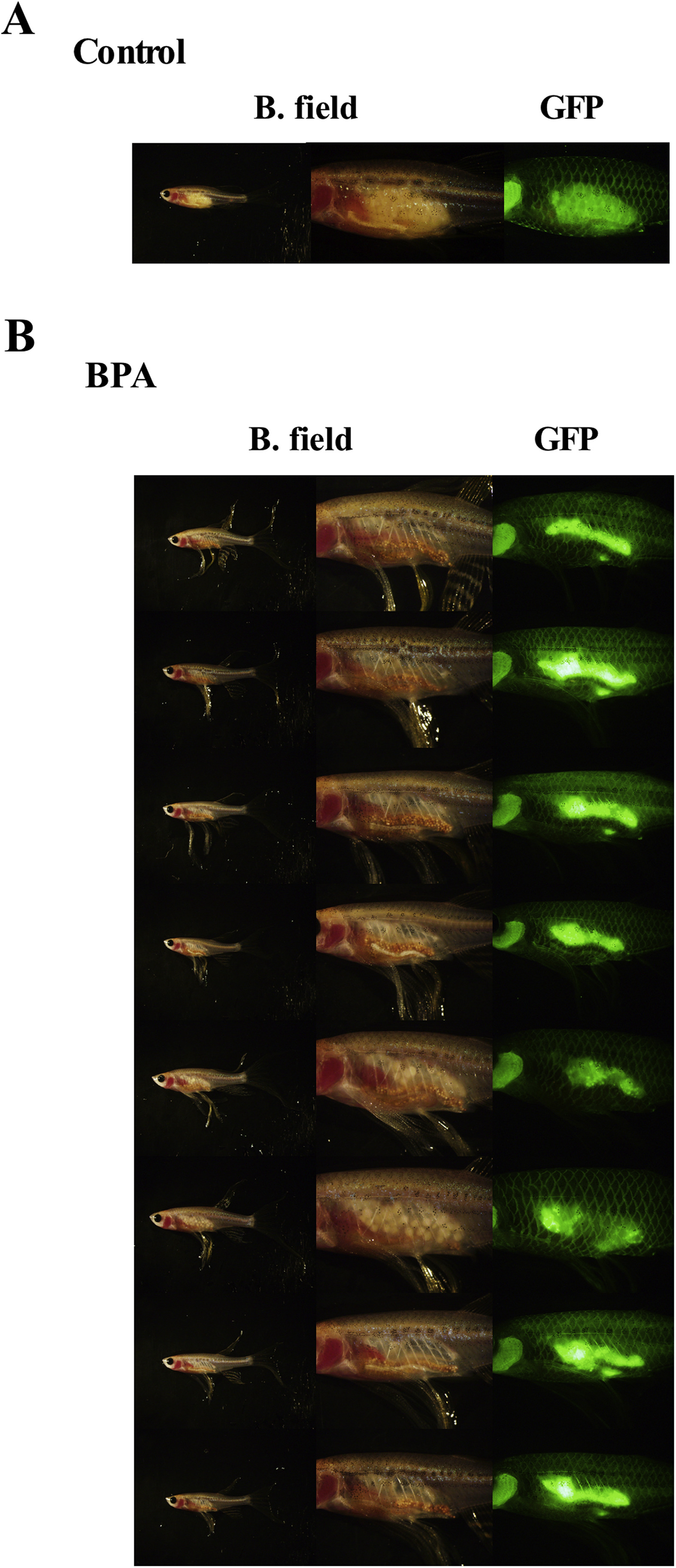
Fig. 4
Adverse effects of BPA treatment on ovaries in the F1 generation of zebrafish. (A) Outside view of a normal β-roy female. (B) Parental male zebrafish were fed BPA-containing food (10 mg of BPA/g food) for 6 months. Then fish were mated with untreated fish (housed separately from experimental fish) and offspring were raised on control food. Outside view of raised female fish generated from BPA-treated males. Note that all indicated fish are seems like males in bright field but they have fluorescent oocytes in abnormal ovaries. Photographs of whole fish and ovarian tissues under bright field (B. field) and GFP filter views (GFP) are shown.