
|
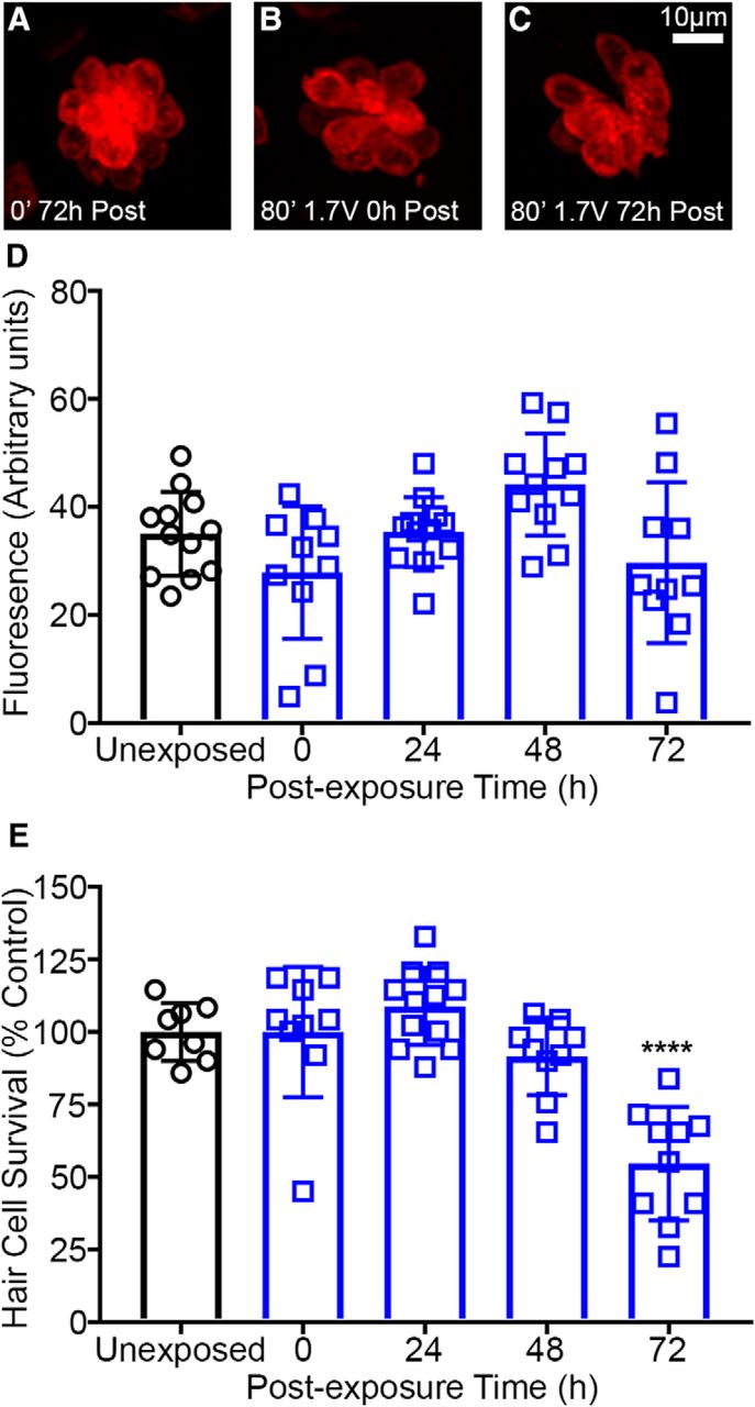
Fig. 5
Loading of the mechanotransduction dependent dye FM 1-43FX is not affected by acoustic stimulation in wild-type *AB zebrafish. A–C, Representative images of neuromasts loaded with FM 1-43FX. Unexposed (A) and acoustically stimulated (B, C) neuromasts are brightly labeled with FM 1-43FX. D, Quantified FM 1-43FX fluorescence (normalized to hair cell number) is not significantly different in unexposed control versus 72 h post-exposure suggesting that acoustic stimulation does not alter hair cell mechanotransduction (one-way ANOVA; post-exposure time: F(4,50) = 4.001, p = 0.0068). Acoustically-exposed fish exhibit highly variable FM 1-43FX loading. E, Hair cell survival is reduced 72 h after acoustic stimulation (one-way ANOVA; post-exposure time: F(4,43) = 17.19, p < 0.0001). Hair cells were labeled with anti-parvalbumin and quantified in fixed animals. Asterisks indicate significant difference from unexposed control (****p < 0.001). N = 8–12 animals per treatment and values represent mean ± SD.