
Fig. 4
- ID
- ZDB-IMAGE-181004-4
- Genes
- Publication
- Wu et al., 2018 - Il34-Csf1r Pathway Regulates the Migration and Colonization of Microglial Precursors
- All Figures
- Figures for Wu et al., 2018
|
Fig. 4
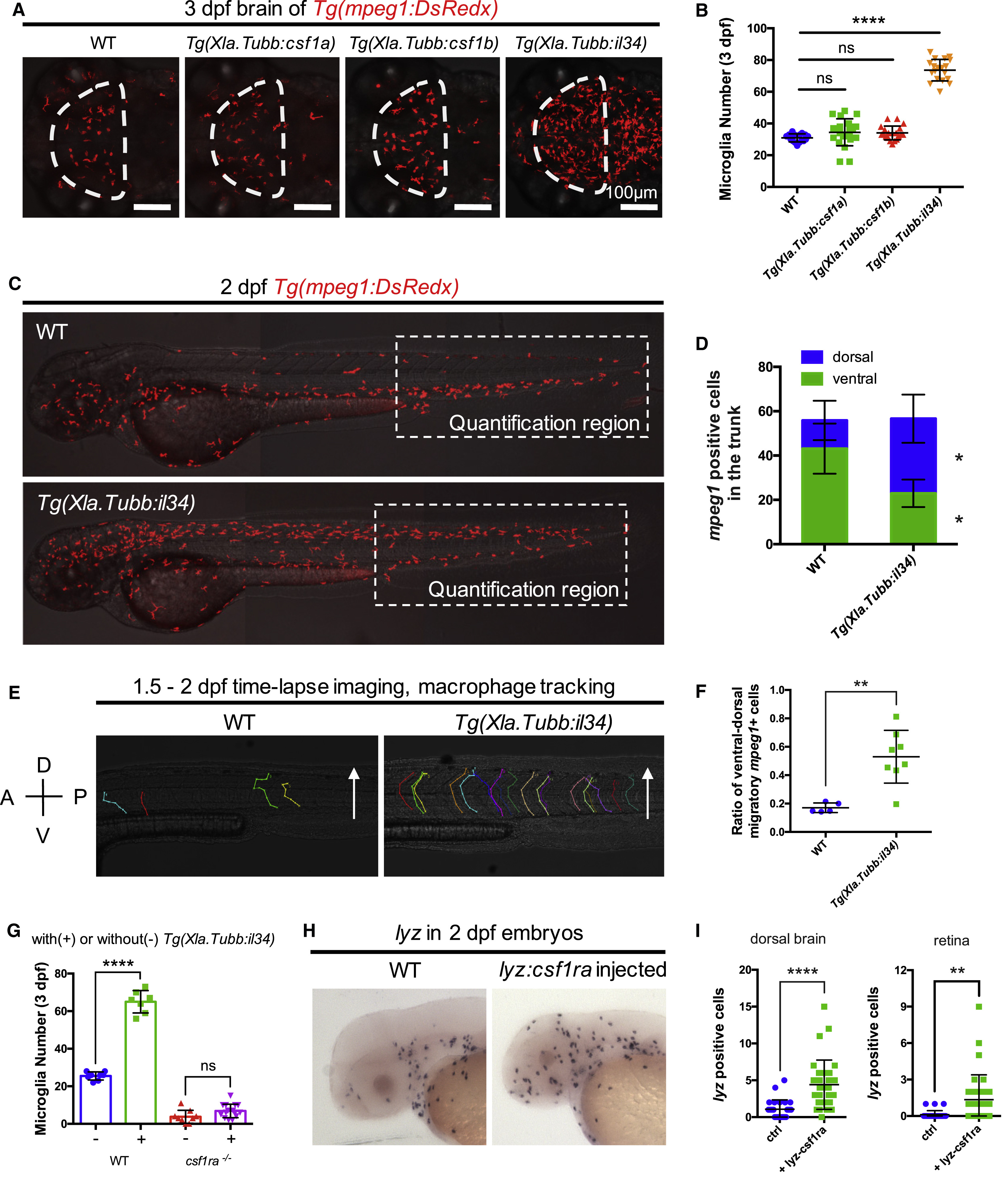
Il34-Csf1ra Pathway Is Sufficient for the Colonization of Microglial Precursors
(A and B) Representative images (A) and quantification (B) of microglia in the optic tectum of 3-dpf WT (n = 17), Tg(Xla.Tubb:csf1a) (n = 22), Tg(Xla.Tubb:csf1b) (n = 20), and Tg(Xla.Tubb:il34) (n = 19) embryos. Microglia were labeled by Tg(mpeg1:DsRedx) (red). The optic tectum is indicated by dashed lines. Values represent means ± SD.
(C and D) Representative images of 2-dpf WT and Tg(Xla.Tubb:il34) embryos showing the distribution of macrophages (C) and quantification of macrophages in the dorsal and ventral region of the trunk in the dashed box region (D). Macrophages were labeled by Tg(mpeg1:DsRedx) (red). n = 14 and 17 for WT and Tg(Xla.Tubb:il34) embryos, respectively. Values represent means ± SD.
(E and F) Time-lapse imaging of the trunk of WT Tg(mpeg1:DsRedx) (n = 5) and Tg(Xla.Tubb:il34;mpeg1:DsRedx) embryos (n = 8) from 36 hpf to 2 dpf, showing the cell tracking (E) and quantification (F) of DsRedx+ macrophages migrating from the ventral trunk region to the dorsal spinal cord region. Values represent means ± SD. A, anterior; P, posterior; D, dorsal; V, ventral. See also Videos S3 and S4.
(G) Quantification of NR+ microglia number in the optic tectum of 3-dpf WT and csf1ra−/− embryos with or without the Tg(Xla.Tubb:il34) transgene. n = 8, 7, 9, and 15 for WT, WT with Tg(Xla.Tubb:il34), csf1ra−/−, and csf1ra−/− with Tg(Xla.Tubb:il34), respectively. Values represent means ± SD. See also Figure S5G.
(H and I) WISH showing lyz expression (H) and quantification of lyz+ neutrophils in the dorsal brain (I, left) and retina (I, right) of 2-dpf WT or lyz:csf1ra-injected embryos. n = 26, 32, 26, and 32 for WT dorsal brain, injected dorsal brain, WT retina, and injected retina, respectively. Values represent means ± SD.
ns, p > 0.05; ∗p ≤ 0.05; ∗∗p ≤ 0.01; ∗∗∗∗p ≤ 0.0001.
Reprinted from Developmental Cell, 46, Wu, S., Xue, R., Hassan, S., Nguyen, T.M.L., Wang, T., Pan, H., Xu, J., Liu, Q., Zhang, W., Wen, Z., Il34-Csf1r Pathway Regulates the Migration and Colonization of Microglial Precursors, 552-563.e4, Copyright (2018) with permission from Elsevier. Full text @ Dev. Cell