
Fig. 1
- ID
- ZDB-IMAGE-181004-1
- Genes
- Publication
- Wu et al., 2018 - Il34-Csf1r Pathway Regulates the Migration and Colonization of Microglial Precursors
- All Figures
- Figures for Wu et al., 2018
|
Fig. 1
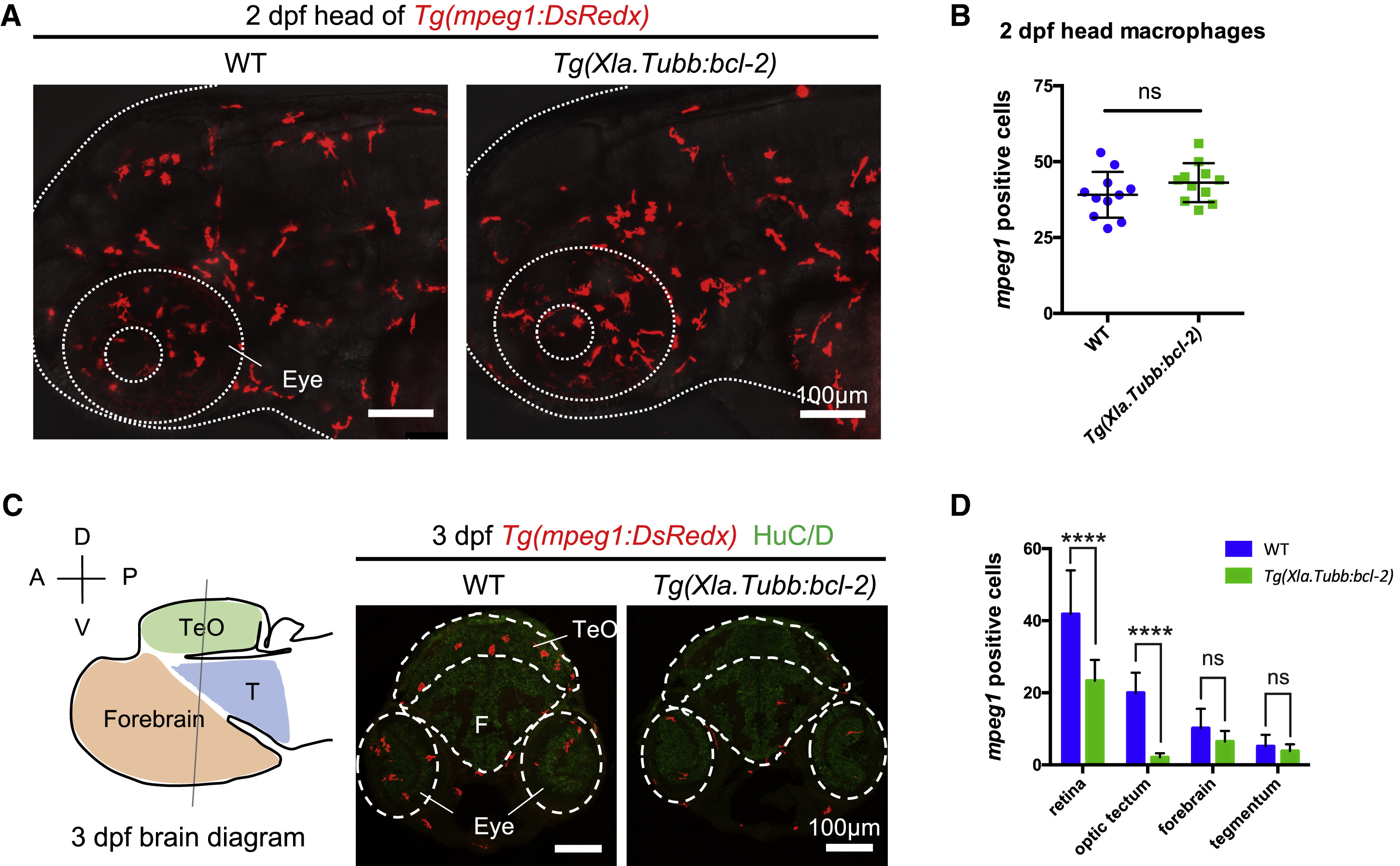
Inhibition of Neuronal Apoptosis Does Not Completely Block Microglia Colonization
(A and B) Lateral view of the head region (A) and quantification (B) of the number of macrophages in the head of 2-dpf WT or Tg(Xla.Tubb:bcl-2) embryos. Macrophages were labeled by Tg(mpeg1:DsRedx) (red). The head is indicated by dotted lines. n = 11 for each group. Values represent means ± SD.
(C) Left: schematic diagram of the 3-dpf zebrafish brain showing the different brain regions. Right: representative transverse sections through the midbrain and hindbrain in 3-dpf WT or Tg(Xla.Tubb:bcl-2) embryos, with macrophages labeled by Tg(mpeg1:DsRedx) (red) and neurons labeled by HuC/D immunostaining (green). Different CNS regions are indicated by dashed lines. The diagram is modified from the Atlas of Early Zebrafish Brain Development: A Tool for Molecular Neurogenetics [M]. Academic Press, 2015. A, anterior; P, posterior; D, dorsal; V, ventral; TeO, optic tectum; T, tegmentum; F, forebrain.
(D) Quantification of DsRedx+ microglia in different CNS regions of 3-dpf WT Tg(mpeg1:DsRedx) or Tg(Xla.Tubb:bcl-2;mpeg1:DsRedx) embryos. n = 11 for each group. Values represent means with SD.
For multiple comparisons, see also Table S1. ns, p > 0.05; ∗∗∗∗p ≤ 0.0001.
Reprinted from Developmental Cell, 46, Wu, S., Xue, R., Hassan, S., Nguyen, T.M.L., Wang, T., Pan, H., Xu, J., Liu, Q., Zhang, W., Wen, Z., Il34-Csf1r Pathway Regulates the Migration and Colonization of Microglial Precursors, 552-563.e4, Copyright (2018) with permission from Elsevier. Full text @ Dev. Cell