
Image
|
Figure Caption
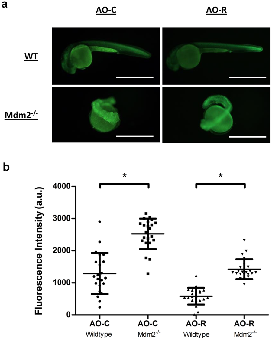
Fig. 6
In-vivo staining of live zebrafish embryos using compound AO-R. (a) Live imaging of either wildtype (top panels), or Mdm−/− (bottom panels) 1 day post fertilization zebrafish embryos stained using either AO-C (left panels) or AO-R (right panels) dyes. Scale bars measure 1 mm. (b) Fluorescence intensity of individually measured whole, wildtype or Mdm2 knock-out, zebrafish embryos stained with either AO-C or AO-R dyes. (N = 22; *P < 0.0001, unpaired, two-tailed student t test).
Acknowledgments
This image is the copyrighted work of the attributed author or publisher, and
ZFIN has permission only to display this image to its users.
Additional permissions should be obtained from the applicable author or publisher of the image.
Full text @ Sci. Rep.