
IMAGE
Fig. 2
- ID
- ZDB-IMAGE-180919-3
- Publication
- Zhang et al., 2018 - Monitoring antiangiogenesis of bevacizumab in zebrafish
- All Figures
- Figures for Zhang et al., 2018
Image
|
Figure Caption
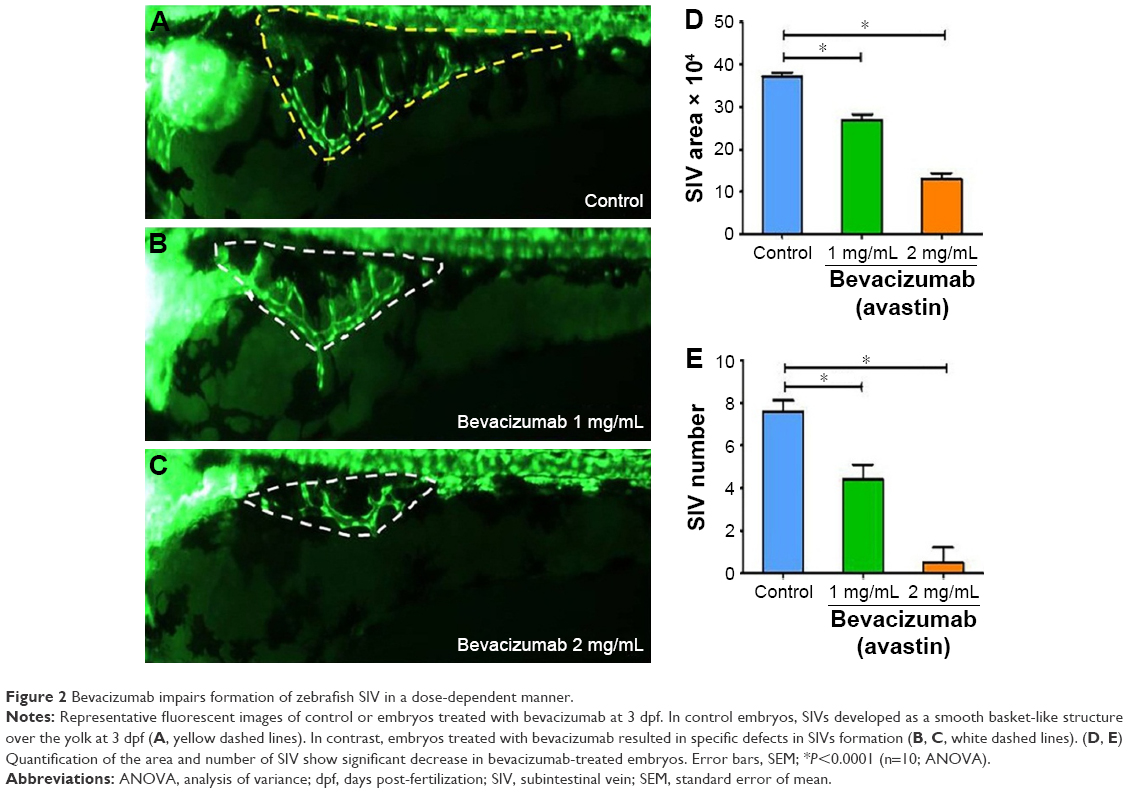
Fig. 2
Bevacizumab impairs formation of zebrafish SIV in a dose-dependent manner.
Representative fluorescent images of control or embryos treated with bevacizumab at 3 dpf. In control embryos, SIVs developed as a smooth basket-like structure over the yolk at 3 dpf (A, yellow dashed lines). In contrast, embryos treated with bevacizumab resulted in specific defects in SIVs formation (B, C, white dashed lines). (D, E) Quantification of the area and number of SIV show significant decrease in bevacizumab-treated embryos. Error bars, SEM; *P<0.0001 (n=10; ANOVA).
Acknowledgments
This image is the copyrighted work of the attributed author or publisher, and
ZFIN has permission only to display this image to its users.
Additional permissions should be obtained from the applicable author or publisher of the image.
Full text @ Drug Des Devel Ther