
IMAGE
Fig. 1
- ID
- ZDB-IMAGE-180919-2
- Publication
- Zhang et al., 2018 - Monitoring antiangiogenesis of bevacizumab in zebrafish
- All Figures
- Figures for Zhang et al., 2018
Image
|
Figure Caption
Fig. 1
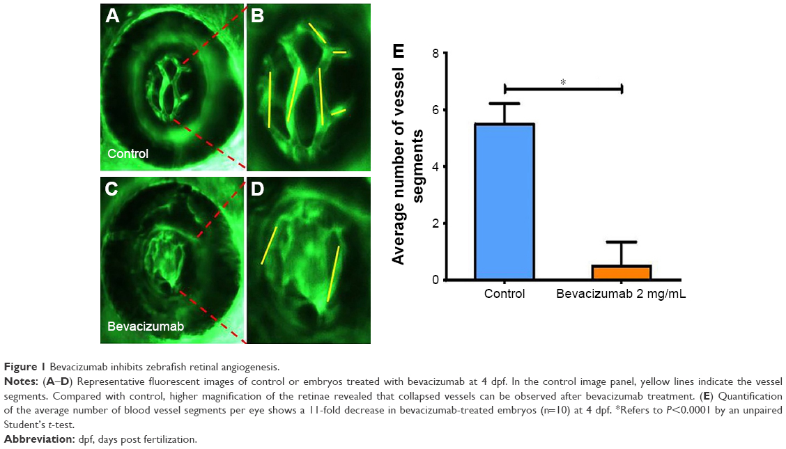
Bevacizumab inhibits zebrafish retinal angiogenesis.
(A–D) Representative fluorescent images of control or embryos treated with bevacizumab at 4 dpf. In the control image panel, yellow lines indicate the vessel segments. Compared with control, higher magnification of the retinae revealed that collapsed vessels can be observed after bevacizumab treatment. (E) Quantification of the average number of blood vessel segments per eye shows a 11-fold decrease in bevacizumab-treated embryos (n=10) at 4 dpf. *Refers to P<0.0001 by an unpaired Student’s t-test.
Acknowledgments
This image is the copyrighted work of the attributed author or publisher, and
ZFIN has permission only to display this image to its users.
Additional permissions should be obtained from the applicable author or publisher of the image.
Full text @ Drug Des Devel Ther