
Fig. 7
- ID
- ZDB-IMAGE-180917-22
- Genes
- Publication
- Svahn et al., 2018 - Nucleo-cytoplasmic transport of TDP-43 studied in real time: impaired microglia function leads to axonal spreading of TDP-43 in degenerating motor neurons
- All Figures
- Figures for Svahn et al., 2018
|
Fig. 7
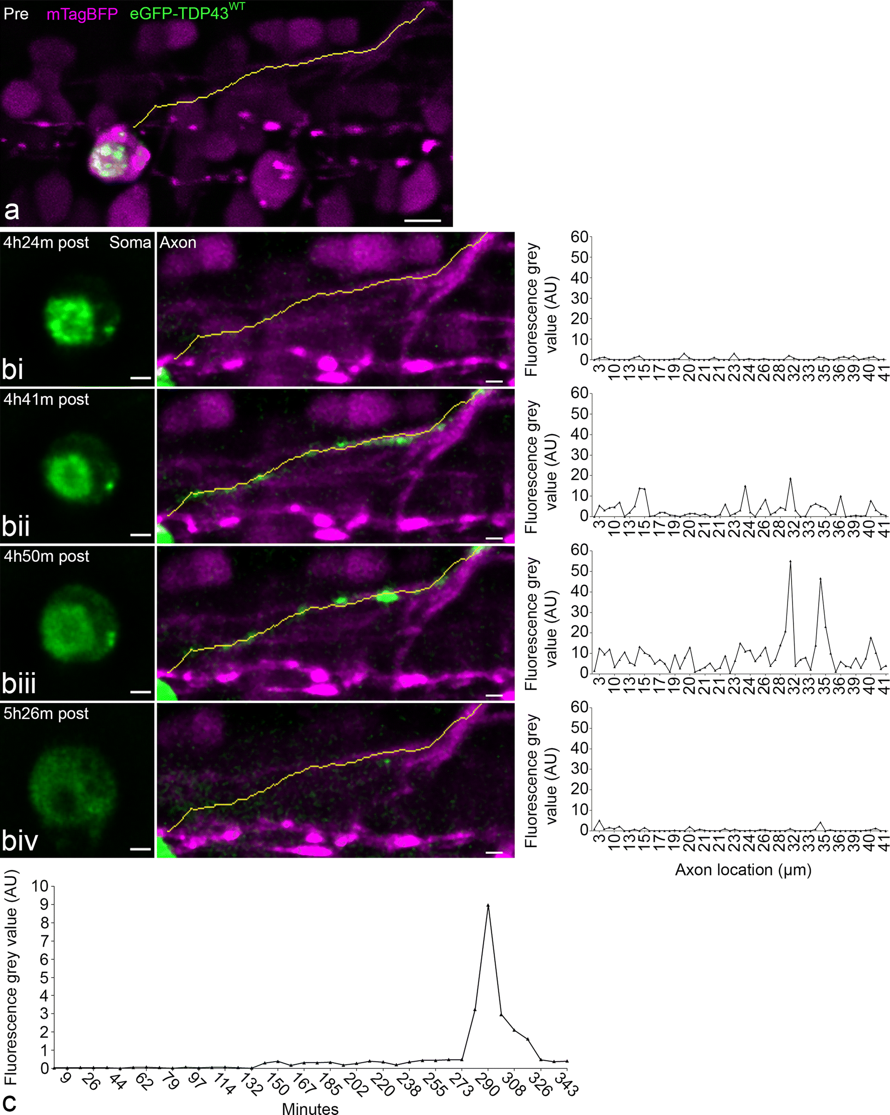
Axonal redistribution of eGFP-TDP43WT during UV-induced degeneration and microglia depletion. a Pre-irradiation, line is a projection of the axon vector used for signal analysis. Scale = 5 µm. bi–iv At each time point from left to right the three windows represents a single time point illustrating the cell body and the TDP-43 distribution (soma, eGFP-TDP43WT in green), the axonal projection (axon, mTagBFP in magenta incl. neurite vector as yellow line), and the fluorescence intensity of eGFP in the segments along the neurite vector. c Line graph demonstrating the time course of the mean eGFP fluorescence intensity along the axon post-stress induction. Scale = 2 µm. Note: axonal redistribution was observed in 2 of the 10 neurons, which were targeted for UV injury and underwent degeneration