
Fig. 3
- ID
- ZDB-IMAGE-180914-15
- Genes
- Antibodies
- Publication
- Rasmussen et al., 2018 - Fish Scales Dictate the Pattern of Adult Skin Innervation and Vascularization
- All Figures
- Figures for Rasmussen et al., 2018
|
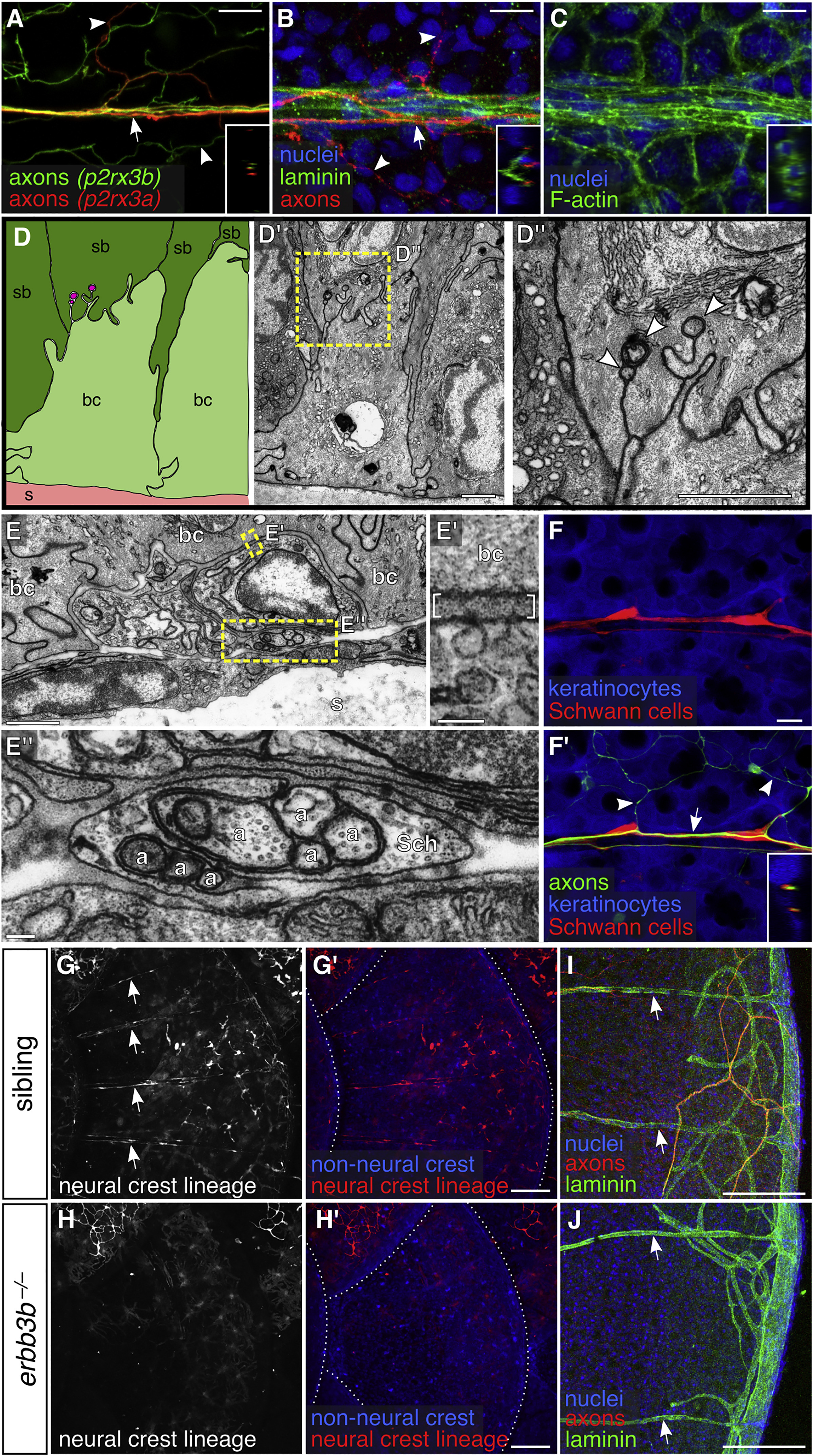
Fig. 3
DRG Axons Form Nerves at the Surface of Scales
(A–C) Projections through a single superficial scale nerve in an adult. Arrow, axon bundle. Arrowheads, axon free endings. Insets, orthogonal views of nerve; outer surface to left.
(D) TEM of scale epidermis. Arrowheads, axon free endings ensheathed by keratinocyte membranes. Yellow box, area of magnification.
(E) Transverse TEM of superficial scale nerve. Yellow boxes, areas of magnification. Brackets in E′ indicate electron dense extracellular material. a, axon; Sch, Schwann cell; bc, basal cell; sb, suprabasal cell; s, scale.
(F) DRG axons and associated Schwann cells (arrow in F′) along the scale surface. Note that free endings in the epidermis (arrowheads) are not associated with Schwann cells. Inset, orthogonal view of nerve.
(G and H) Representative lateral views of the trunk of adult erbb3b−/− and sibling controls. Arrows, scale nerve-associated Schwann cells. Dashed lines, scale margins.
(I and J) Representative scales from adult erbb3b−/− and sibling controls immunostained for indicated markers. Arrows, superficial laminin tracts.
Transgenes: (A) Tg(p2rx3b:EGFP) and Tg(p2rx3a>mCherry) (these two transgenes label nociceptive sensory neurons, but, as in larvae, P2rx3a appears to label a subset of P2rx3b neurons; Gau et al., 2013, Palanca et al., 2013); (F) axons (Tg(p2rx3b:EGFP)), and keratinocytes and Schwann cells (Tg(-28.5Sox10:Cre);Tg(actb2:BswitchR)); (G and H) Tg(-28.5Sox10:Cre);Tg(actb2:BswitchR). Staining: (B, I, and J) laminin (anti-laminin), axons (acTubulin), and nuclei (DAPI); (C) F-actin (phalloidin) and nuclei (DAPI). Scale bars, 10 μm (A–C and F), 1 μm (D and E), 0.1 μm (E′ and E″), and 100 μm (G–J).
Reprinted from Developmental Cell, 46(3), Rasmussen, J.P., Vo, N.T., Sagasti, A., Fish Scales Dictate the Pattern of Adult Skin Innervation and Vascularization, 344-359.e4, Copyright (2018) with permission from Elsevier. Full text @ Dev. Cell