
Fig. S3
- ID
- ZDB-IMAGE-180913-51
- Publication
- Asakawa et al., 2018 - Protocadherin-Mediated Cell Repulsion Controls the Central Topography and Efferent Projections of the Abducens Nucleus
- All Figures
- Figures for Asakawa et al., 2018
|
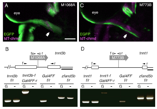
Fig. S3
Compartmental organization of the lateral rectus muscle characterized by troponins. (Related to Figure 1)
(A) Dorsal view of the left lateral rectus muscle in a Tg[gSAIzGFFM1068A]; Tg[UAS:GFP]; Tg[actc1b:tdT-chrnd] larva at 5dpf. The double arrowheads indicate the presumptive initial contact sites between the abducens motor neurons and lateral rectus muscle. Rostral is to the top. (B) Tg[gSAIzGFFM1068A], indicated as M1068A, traps tnnt3b. (C) Dorsal view of the left lateral rectus muscle in a Tg[gSAIzGFF773B]; Tg[UAS:EGFP]; Tg[actc1b:tdT-chrnd] larva at 5dpf. (D) Tg[gSAIzGFF773B], indicated as M773B, traps tnnt1. The open boxes represent exons of the troponin genes. Chimeric transcript between the troponin genes and Gal4 was detectable by RT-PCR (bottom). The filled and open arrowheads show the position and direction (f: forward, and r: reverse) of primers annealing with the troponin exons and Gal4, respectively. For each set of primer pairs, cDNA reverse-transcribed from poly A-tailed RNA of fish with (G, left lane) or without (-, right lane) the gene trap construct was examined. The ubiquitously expressed zfand5b was used as a control. The bars indicate 50 μm in A, C and 100 bp in B, D.