
|
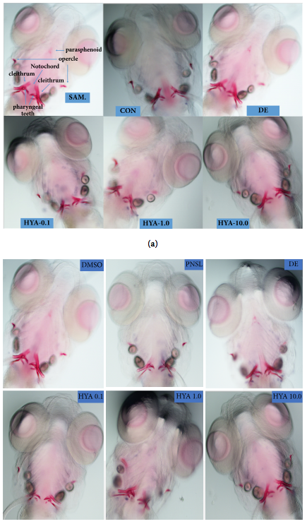
Fig. 1
Ventral view of Alizarin Red stained zebrafish skull at 10 dpf (×100). (a) Exposure to blank E3 medium and the effect of HYA on bone mineralization. SAM, the structure of zebrafish bones after staining; CON, blank E3 medium; DE, 15 μM disodium ethydronate; HYA 0.1, 0.1 μM HYA; HYA 1.0, 1.0 μM HYA; HYA 10.0, 10.0 μM HYA. (b) Exposure to prednisolone and the therapeutic effect of HYA on GCIOP. CON, blank E3 medium; DMSO, blank E3 medium + 0.5% DMSO; PNSL, blank E3 medium + 0.5% DMSO + 25 μM prednisolone; DE, 15 μM disodium ethydronate + PNSL; HYA 0.1, 0.1 μM HYA + PNSL; HYA 1.0, 1.0 μM HYA + PNSL; HYA 10.0, 10.0 μM HYA + PNSL. Note: areas of calcified matrix in craniofacial skeleton are stained red.