
|
Fig. 1
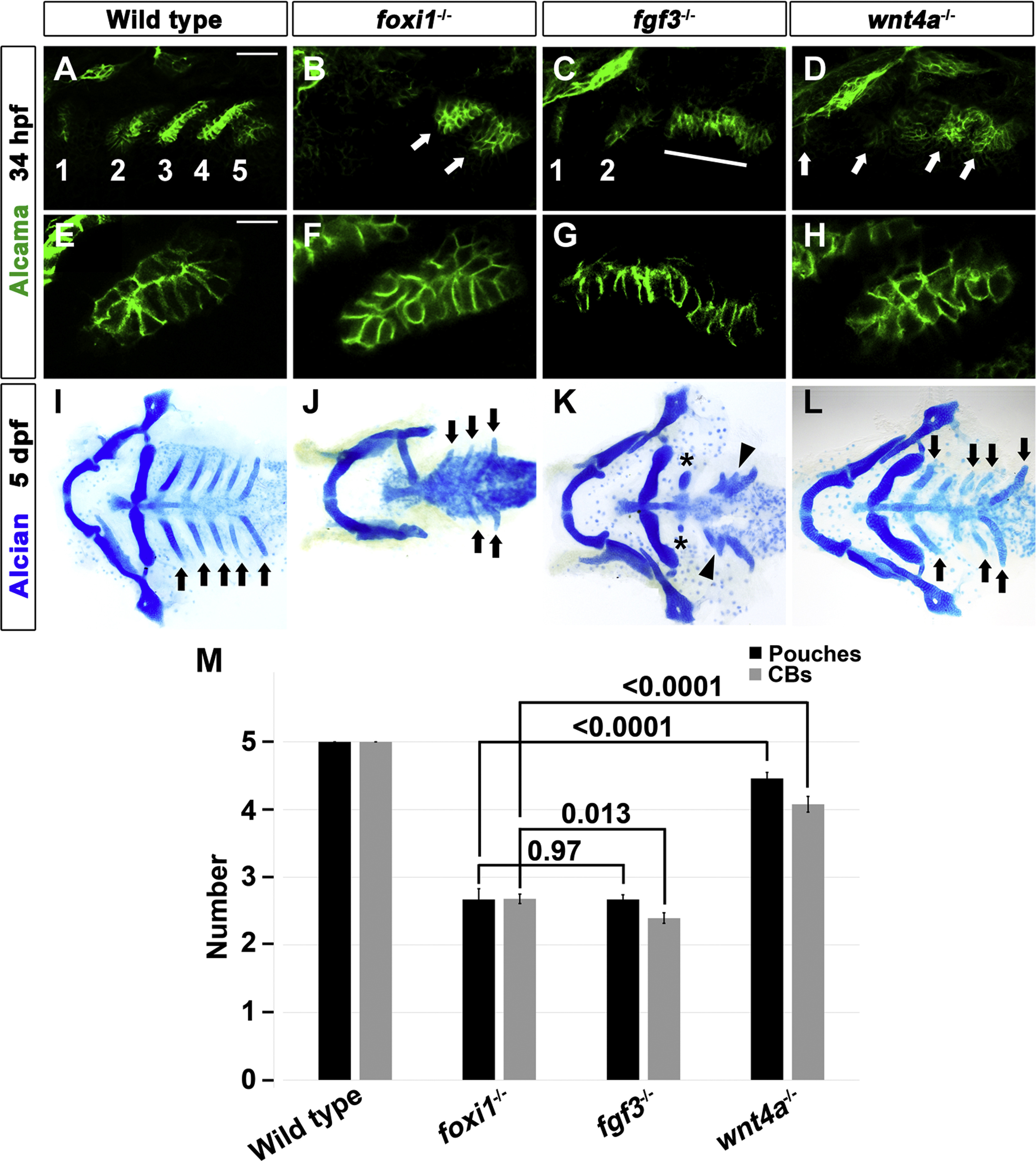
Requirement for Foxi1 in the late-stage of pouch development. (A-H) Alcama immunohistochemistry (green) showed five pouches (1–5 in A) in wild-type embryos at 34 hpf. foxi1 mutants had posterior pouches shaped abnormally (arrows in B), whereas fgf3 mutants had two anterior pouches (1 and 2 in C) with a cell mass that failed to migrate out to form posterior pouches (line in C). wnt4a mutants displayed four abnormally shaped pouches (arrows in D). Scale bar: 40 µm (A-D). (E) Alcama immunohistochemistry (green) showed a normal bilayered pouch morphology in wild-type embryos. (F and H) In foxi1 and wnt4a mutants, an aberrant, similar multilayered pouch morphology was found. (G) In fgf3 mutants, posterior pouch-forming cells failed to migrate out. Scale bar: 20 µm (E-H). (I-L) Ventral views of facial cartilages. In wild-type embryos, a bilateral set of five CBs (arrows in I) formed. Fewer CBs were observed in foxi1 and wnt4a mutants, whereas fusions of CBs (arrowheads in K) were observed with reduced anterior CBs (asterisks in K) in fgf3 mutants. (J, L) Normally shaped CBs were marked with arrows in foxi1 and wnt4a mutants. (M) Quantification of pouch and CB defects in wild-type embryos and mutants. Data represent mean ± s.e.m. P values are shown.
Reprinted from Developmental Biology, 441(1), Jin, S., Jiyun, O., Stellabotte, F., Choe, C.P., Foxi1 promotes late-stage pharyngeal pouch morphogenesis through ectodermal Wnt4a activation, 12-18, Copyright (2018) with permission from Elsevier. Full text @ Dev. Biol.