
Fig. 6
- ID
- ZDB-IMAGE-180906-40
- Publication
- Ouyang et al., 2017 - Hyaluronic acid synthesis is required for zebrafish tail fin regeneration
- All Figures
- Figures for Ouyang et al., 2017
|
Fig. 6
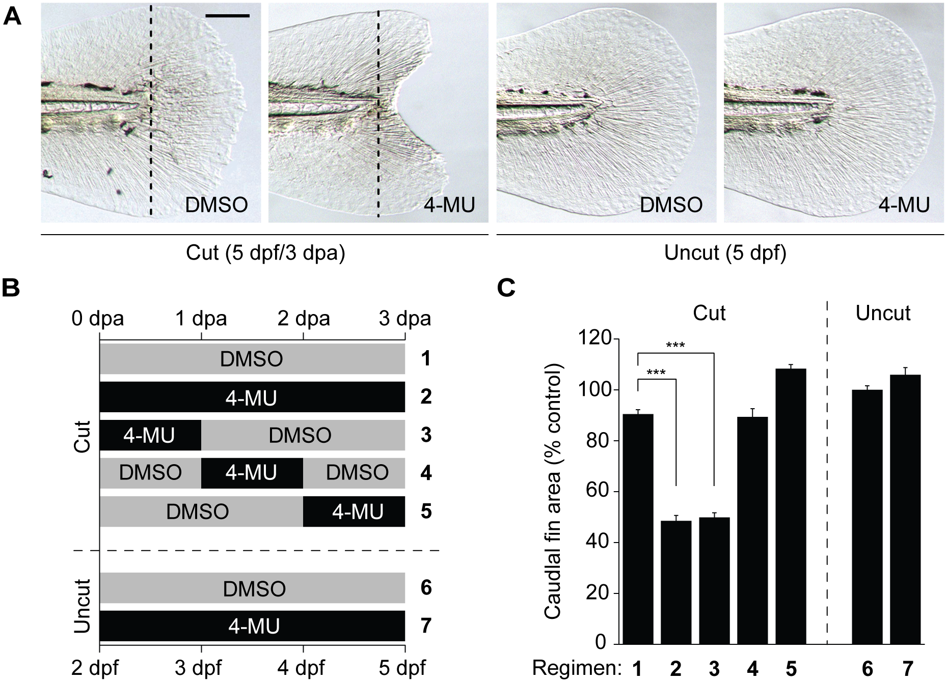
4-MU inhibits larval tail regeneration.
(A) Representative micrographs of larval tails that were amputated at 2 dpf and then treated 0.5% DMSO or 150 μM 4-MU for 3 days. Dotted lines indicate the amputation plane, and micrographs of uncut larval tails subjected to the same inhibitor regimen are shown for comparison. Scale bar: 100 μm. (B-C) Time-course analysis of 4-MU action on larval tail regeneration. Caudal fin sizes at 5 dpf (3 dpa) after the indicated amputation and 4-MU treatment regimens. Data are the average caudal fin areas of 15 larvae ± s.e.m., normalized to the average fin size of uncut larvae treated with 0.5% DMSO. ***, P < 0.001.