
|
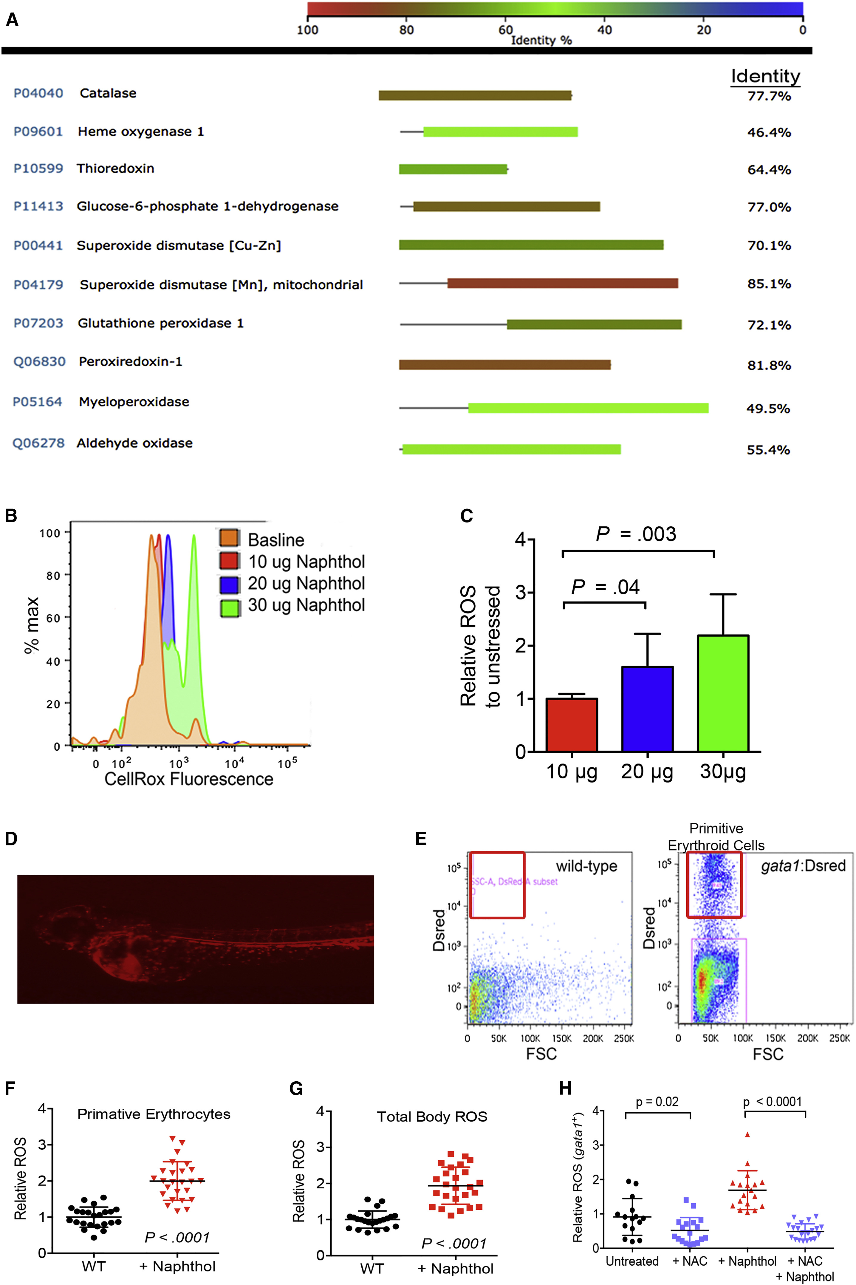
Fig. 1
Oxidative Stress in gata1+ Erythroid Cells
(A) BLASTP identity analysis of key proteins involved in the oxidative stress response between Danio rerio and Homo sapiens.
(B) Representative flow cytometry histograms showing CellRox emission in 72 hpf embryos after 48 hr of 1-naphthol exposure (concentrations shown as μg per 5 mL).
(C) Quantification of CellRox probe signal MFI relative to unstressed embryos. n = 20 individual animals per condition showing one of five independent experiments.
(D) Live imaging of a gata1:DsRed transgenic zebrafish at 72 hpf indicating DsRed-positive primitive erythroid cells.
(E) Representative flow cytometry of single-cell suspensions prepared from wild-type and gata1:DsRed zebrafish at 72 hpf.
(F and G) Pro-oxidant exposure induces ROS in Gata1+ erythroid precursors. Animals were exposed to 20 μg/5 mL 1-naphthol followed by flow cytometry of Gata1+ erythroid cells by gating on DsRed-positive cells (or all cells for total-body ROS) and measuring CellRox probe MFI to determine ROS. n = 20–25 individual animals per condition showing one of four independent experiments.
(H) NAC combined with 1-naphthol reduces ROS in Gata1+ erythroid cells. All pro-oxidant exposure times were from 24 to 72 hpf.
All data are shown as the mean ± SD, with the p value from a Student t test. See also Figure S1.