
|
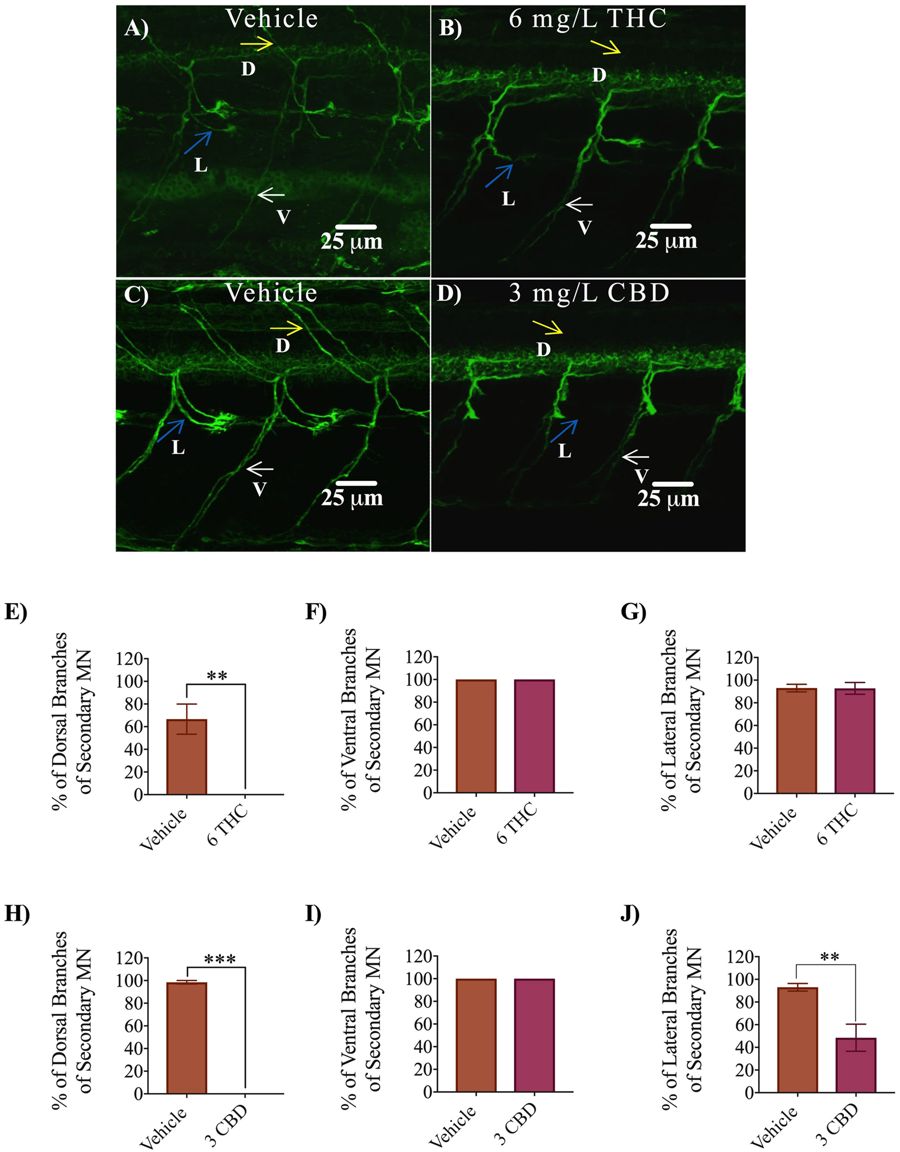
Fig. 6
Antibody labelling (anti-zn8) of axonal branches of secondary motor neurons in 2 dpf embryos in vehicle control, 6 mg/L THC-treated embryos and 3 mg/L CBD treated embryos. (A–D) Dorsal, ventral and lateral branches emanating from secondary motor neurons are indicated by yellow, white and blue arrows. Dorsal branches were absent in THC and CBD treated embryos (B,D). Fewer lateral branches are visible in CBD treated embryos. (E–G) Bar graph comparing percentage of dorsal branches (E), ventral branches (F) and lateral branches (G) emanating from secondary motor neurons in vehicle control (n = 11) and 6 mg/L THC treated embryos (n = 11). (H–J) Bar graph comparing percentage of dorsal branches (H), ventral branches (I) and lateral branches (J) emanating from secondary motor neurons in vehicle control (n = 11) and 3 mg/L CBD treated embryos (n = 9). **Significantly different from vehicle control, p < 0.01. ***Significantly different from vehicle control, p < 0.001.