
|
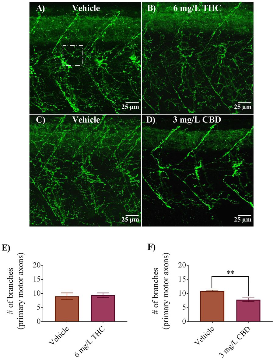
Fig. 5
Antibody labelling (anti-znp1) of axonal branches of primary motor neurons in 2 dpf embryos in vehicle controls, 6 mg/L THC-treated embryos and 3 mg/L CBD treated embryos. (A–D) Branching patterns and labelling of axons appear to be similar between controls and THC-treated embryos but reduced in CBD treated embryos. (E) Bar graph showing the number of branches emanating from primary motor axons in vehicle control (n = 7) and 6 mg/L THC treated embryos (n = 8), counted from 9 different square areas (each 1500 μm2 area). (F) Bar graph showing the number of branches emanating from primary motor axons in vehicle control (n = 6) and 3 mg/L CBD treated embryos (n = 8), counted from 9 different square areas (each about 1500 μm2 area). *Significantly different from vehicle control, p < 0.01.