
Fig. 4
- ID
- ZDB-IMAGE-180828-38
- Publication
- Phan et al., 2018 - Neutrophils use superoxide to control bacterial infection at a distance
- All Figures
- Figures for Phan et al., 2018
|
Fig. 4
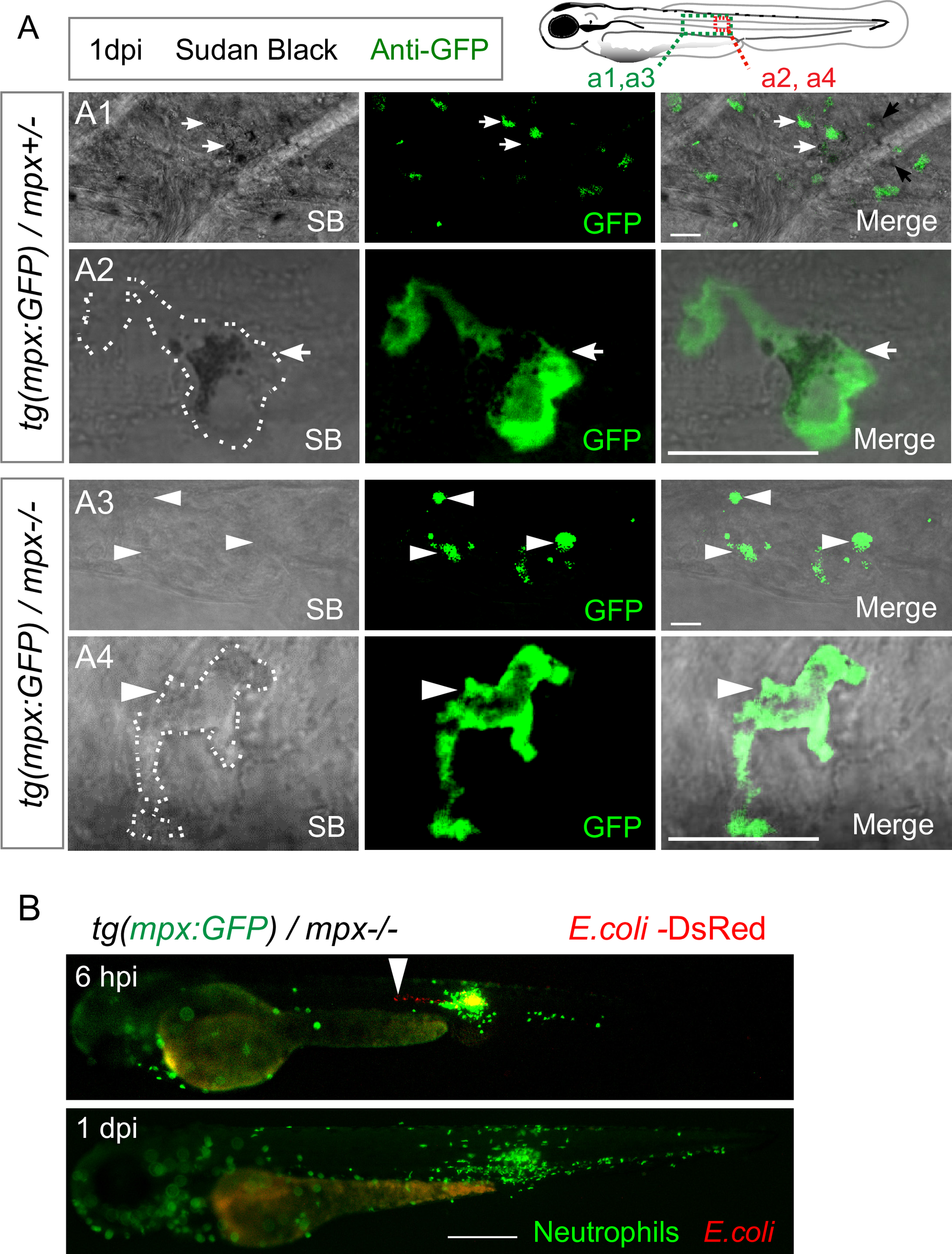
MPO is not required for bacterial clearance in the notochord.
(A) Tg(mpx:GFP)/mpx+/- (A1, A2) and tg(mpx:GFP)/mpx-/- (A3, A4) embryos were infected with E. coli in the notochord. Sudan Black staining and immuno-detection of neutrophils (anti-GFP) were performed in whole embryos at 1 dpi. The top right panel shows the regions imaged by confocal microscopy in the larvae in A1 and A3 (green box) and in A2 and A4 (red box). Representative transmitted light images, overlaid with a maximal projection of confocal fluorescence images show the presence of black granules in the neutrophils (white arrows) of tg(mpx:GFP)/mpx+/- embryos. MPX granules are absent in neutrophils (white arrowheads) of tg(mpx:GFP)/mpx-/- embryos. Scale bars: 10 μm and white dotted lines outline neutrophils. (B) Tg(mpx:GFP)/mpx-/- embryos were infected with E. coli-DsRed in the notochord. Neutrophils (GFP) and E. coli (DsRed) were imaged repeatedly in individual larvae using fluorescent microscopy at 6 hpi and 1 dpi. While E. coli locates in the notochord at 6 hpi (arrowheads), it disappears at 1 dpi. (Nmpx+/- = 9, Nmpx-/- = 8 embryos per condition, from two independent experiments). Scale bar: 400 μm.