
Image
|
Figure Caption
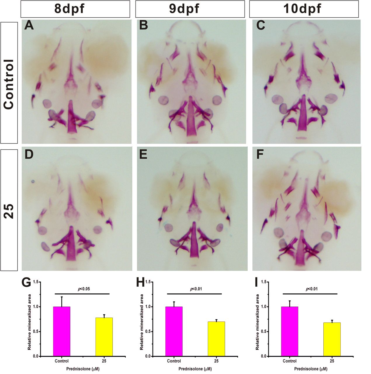
Fig. 1
Establishment of a zebrafish larva GIOP model using 25 μM prednisolone. (A-F) 8−10 dpf zebrafish treated with 25 μM prednisolone. Whole mount skeletal staining was performed on fixed tissues. The mineralized tissue is stained purple and all other tissues are transparent. (G-I) Digital image analysis of stained area and staining density by assessment of fluorescence intensity. The stained mineralized tissue was quantified using analysis software. Mean values are plotted (n=10) and the Student's t-test was performed to determine statistical significance.
Figure Data
Acknowledgments
This image is the copyrighted work of the attributed author or publisher, and
ZFIN has permission only to display this image to its users.
Additional permissions should be obtained from the applicable author or publisher of the image.
Full text @ Biol. Open