
|
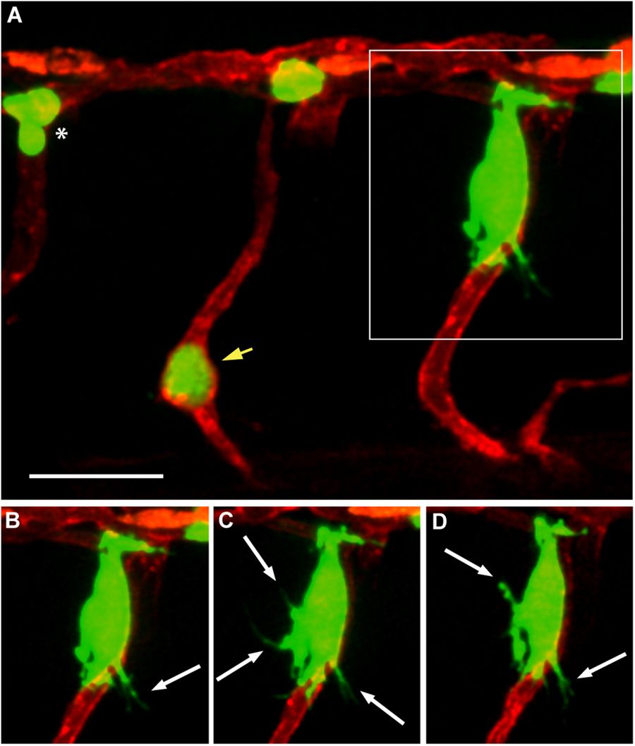
Fig. 6
Angiotropism in zebrafish xenograft of uveal melanoma. (A) A larva injected with OMM 2.3-GFP cells, displaying a micrometastasis of angiotropic cells (in the square) cuffing the external surface of an intersegmental vessel. (B–D) are time-lapse images of the same angiotropic cells taken at time 0, 4 and 8 hours after the beginning of the imaging. The images were obtained employing a Zeiss LSM 880 confocal microscope (40 × water objective), starting from 30 hours post injection. Scale bar is 20 µm, green shows melanoma cells, red shows zebrafish blood vessels, white arrows show pseudopodial protrusions formed by angiotropic cells, white asterisk shows intravascular melanoma cells, yellow arrow shows melanoma cells trapped in an intersegmental vessel.