
Fig. S1
- ID
- ZDB-IMAGE-180828-23
- Publication
- Gauvrit et al., 2018 - HHEX is a transcriptional regulator of the VEGFC/FLT4/PROX1 signaling axis during vascular development
- All Figures
- Figures for Gauvrit et al., 2018
|
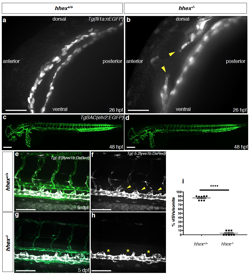
Fig. S1
zebrafish hhex mutants exhibit a delay during PHBC formation but form arterial intersegmental vessels
(a-b) Maximum intensity projections of confocal images of 24 hpf Tg(fli:nEGFP); hhex+/+ and hhex-/- embryos after GFP immunostaining. hhex mutants exhibit delayed primordial hindbrain channels (PHBCs) formation (arrowheads). (c-d) Maximum intensity projections of confocal images of 48 hpf TgBAC(etv2:EGFP); hhex+/+ and hhex-/- embryos. hhex mutants form arterial ISVs. (e-h) Trunk vasculature of 5 dpf Tg(kdrl:EGFP); Tg(-5.2lyve1b:DsRed); hhex+/+ and hhex-/-. hhex mutants form arterial ISVs while vessels from the PCV (vISVs and TD) are mostly absent (arrowheads point to the ventrally positioned TD; asterisks indicate lack of this structure). i) Quantification of vISVs across 10 somites in 5 dpf hhex+/? (n=8) and hhex-/- (n=8). Values represent means ± s.e.m. ****P ≤0.0001 by t-test. Scale bars: 100 μm (a-b), 200 μm (c-d), 50 μm (e-h).