
IMAGE
Fig. 5
- ID
- ZDB-IMAGE-180827-79
- Publication
- Tseng et al., 2018 - Modeling Niemann-Pick disease type C1 in zebrafish: a robust platform for in vivo screening of candidate therapeutic compounds.
- All Figures
- Figures for Tseng et al., 2018
Image
|
Figure Caption
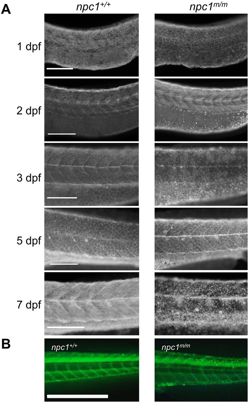
Fig. 5
Unesterified cholesterol accumulation. (A) Control and mutant larvae were stained with filipin to visualize unesterified cholesterol. Filipin-positive puncta were initially observed in mutant larvae at 2 dpf in the area of the yolk sac extension and became more numerous with a wider distribution in older larvae. Scale bars: 200 µm. (B) Embryos were injected with TopFluor-cholesterol at the 1-cell stage and imaged at 7 dpf. Puncta of accumulated cholesterol were observed in the npc1m/m larvae. Scale bar: 1 mm.
Figure Data
Acknowledgments
This image is the copyrighted work of the attributed author or publisher, and
ZFIN has permission only to display this image to its users.
Additional permissions should be obtained from the applicable author or publisher of the image.
Full text @ Dis. Model. Mech.