
IMAGE
Fig. 3
- ID
- ZDB-IMAGE-180827-77
- Publication
- Tseng et al., 2018 - Modeling Niemann-Pick disease type C1 in zebrafish: a robust platform for in vivo screening of candidate therapeutic compounds.
- All Figures
- Figures for Tseng et al., 2018
Image
|
Figure Caption
Fig. 3
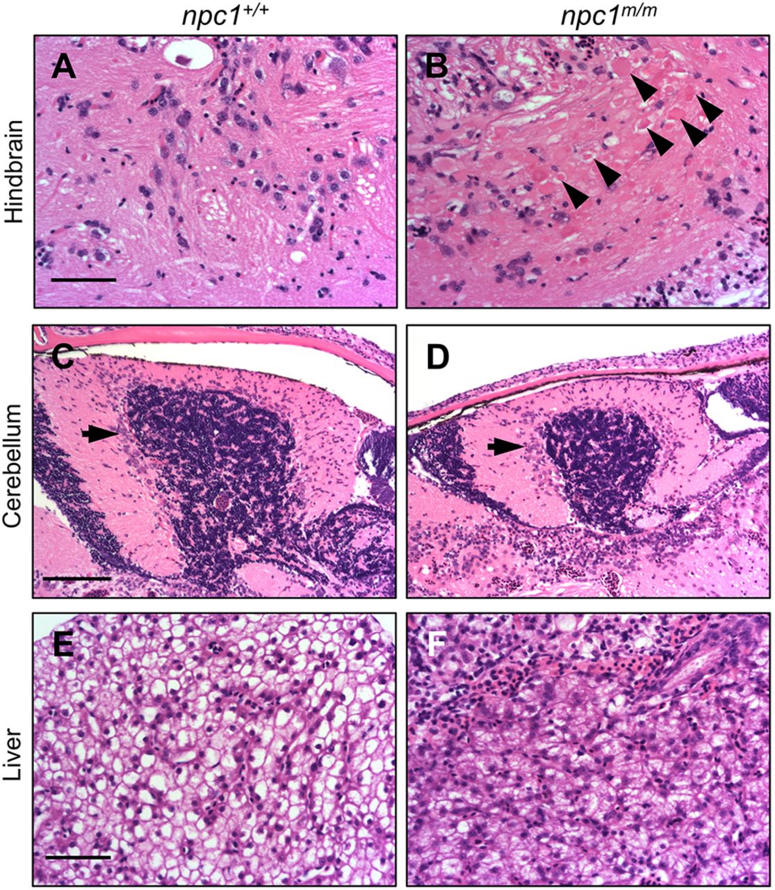
Histopathology of adult npc1m/m liver and brain tissue. (A,B) Photographs of Hematoxylin and Eosin (H&E)-stained hindbrain tissue sections from adult npc1+/+ (A) and npc1m/m (B) zebrafish at 9 weeks of age. Axonal spheroids are indicated by the arrowheads. Scale bar: 50 μm. (C,D) H&E-stained cerebellar sections from 9-week npc1+/+ (C) and npc1m/m (D) zebrafish. The arrows indicate cerebellar Purkinje neurons. Scale bar: 100 μm. (E,F) H&E-stained liver sections from 9-week npc1+/+ (E) and npc1m/m (F) zebrafish. Scale bar: 50 μm.
Figure Data
Acknowledgments
This image is the copyrighted work of the attributed author or publisher, and
ZFIN has permission only to display this image to its users.
Additional permissions should be obtained from the applicable author or publisher of the image.
Full text @ Dis. Model. Mech.