Fig. S1
Genes, Phenotypes, and Scoring Conventions. (Related to Figures 1-6)
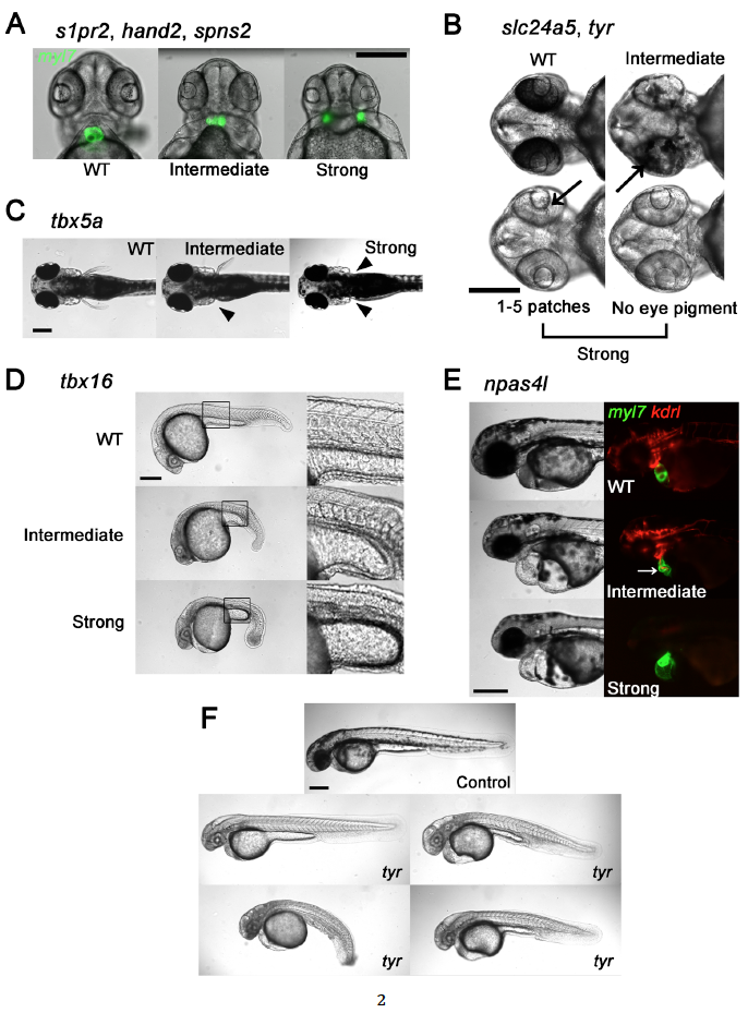
Phenotypes that mimicked those seen in nulls produced by heterozygous mutant lines were scored as Strong and less severe phenotypes were scored as Intermediate as follows: (A) s1pr2, hand2, and spns2. Heart fields marked by Tg(myl7:GFP), showing normal cardiac morphology (WT), partially-fused and non-functional heart (Intermediate), and cardia bifida (Strong). A Strong cardia bifida phenotype was called if the heart fields were completely separated, independent of their distance. (B) slc24a5 and tyr. Black arrows point to patches of pigmented retinal epithelium. No eye pigment or fewer than 5 small patches was classified as a Strong phenotype. Reduced pigmentation but not Strong was classified as Intermediate. (C) tbx5a. Absence of pectoral fin(s) denoted by solid arrowhead(s). Absence of both fins was classified as a Strong phenotype. Absence of one was Intermediate. (D) tbx16. Intact somite formation with abnormal tail bud formation was classified as Intermediate. phenotype. Lack of somite formation and abnormal tail bud formation was classified as Strong phenotype. (E) npas4l. Embryos with cardiac labeling by Tg(myl7:GFP) and endothelial/endocardial labeling by Tg(kdrl:rasCherry) were used. A Strong phenotype was defined as a cloche (bell) heart and absence of endothelial marker. A non-functional misshapen heart (white arrow) with partial endothelial/endocardial labeling was classified as Intermediate. Scale bars, 250 μm. (F) Spectrum of body morphologies in embryos injected with Cas9 RNP with 16 guides directed at the tyr allele. Note embryo with grossly normal body shape as upper left and three dysmorphic embryos with curvature or gross tail, trunk or yolk malformations. Scale bar 250 μm for all images.
Reprinted from Developmental Cell, 46, Wu, R.S., Lam, I.I., Clay, H., Duong, D.N., Deo, R.C., Coughlin, S.R., A Rapid Method for Directed Gene Knockout for Screening in G0 Zebrafish, 112-125.e4, Copyright (2018) with permission from Elsevier. Full text @ Dev. Cell

