
Fig. S1
- ID
- ZDB-IMAGE-180822-58
- Publication
- Gribble et al., 2018 - The synaptic receptor Lrp4 promotes peripheral nerve regeneration
- All Figures
- Figures for Gribble et al., 2018
|
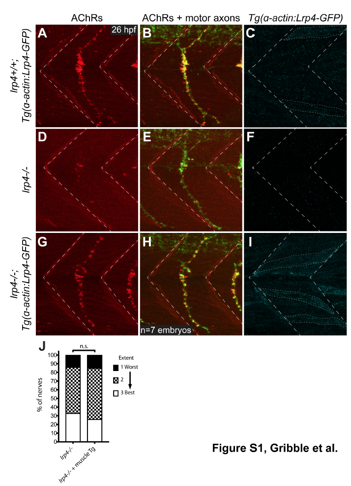
Fig. S1
Selective expression of lrp4 in muscle cells rescues neuromuscular synaptic defects in lrp4 mutants (A) Lateral view of single hemisegment in a 26 hpf wild-type zebrafish embryo expressing Tg(α-actin:Lrp4-GFP), showing normal distribution of acetylcholine receptors (AChRs) in the center of the hemisegment. (B) Merged image of AChRs and motor axons labeled with znp-1 antibody, showing AChRs clustered beneath motor axon terminals. (C) Localization and expression of Tg(α-actin:Lrp4-GFP) in wild-type muscle cells. Lrp4-GFP localizes in puncta distributed throughout each muscle cell. (D,E) lrp4 mutants at 26 hpf show a dramatic reduction in AChR clusters and neuromuscular synapses, but motor axons extend normally. (F) No Lrp4-GFP signal is observed in this mutant, and the muscle rescue transgene is not detectable by PCR. (G,H) lrp4 mutants expressing Tg(α- actin:Lrp4-GFP) show AChR clusters and neuromuscular synapses at 26 hpf that appear wild-type (n=7 embryos). (I) Expression of Tg(α-actin:Lrp4-GFP) in muscle cells of lrp4 mutant embryo. (J) Quantification of nerve regeneration in lrp4 mutant larvae and lrp4 mutant larvae expressing the muscle-specific α-actin:Lrp4-GFP transgene. At 48 hpt, lrp4 mutant larvae expressing Tg(α-actin:Lrp4-GFP) regenerate poorly (n=27 nerves), like lrp4 mutant larvae (n=21 nerves; Chi-square test p=0.8505; Chi-square=0.3238, df=2).