
Fig. 1-S2
- ID
- ZDB-IMAGE-180821-38
- Publication
- Fukui et al., 2018 - Hippo signaling determines the number of venous pole cells that originate from the anterior lateral plate mesoderm in zebrafish
- All Figures
- Figures for Fukui et al., 2018
|
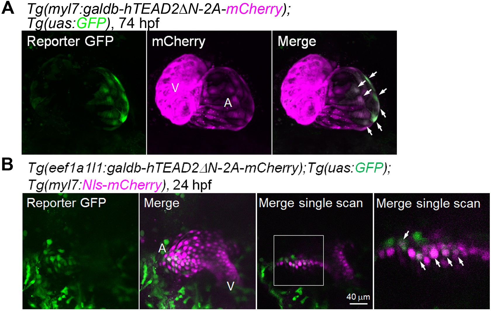
Fig. 1-S2
Tead reporter expression is found in the venous pole CMs of the atrium.
(A) Confocal 3D-stack images (at 74 hpf) of Tg(myl7:galdb-hTEAD2ΔN-2A-mCherry);Tg(uas:GFP) embryos (n = 6). Arrows indicate the Tead reporter GFP-positive atrial CMs. Reporter GFP image (left), mCherry image (center), and merged image of GFP and mCherry (right). A, atrium; V, ventricle. Ventral view, anterior to the top. (B) Confocal images (at 24 hpf) of the Tg(eef1a1l1:galdb-hTEAD2ΔN-2A-mCherry);Tg(uas:GFP);Tg(myl7:Nls-mCherry) embryos (n = 12). The boxed region is enlarged and shown in the panel furthest to the right. Arrows indicate the Tead reporter-positive cells that might differentiate into atrial CMs. Regions in the cardiac tube that would give rise to the atrium (A) and ventricle (V) are marked. Confocal 3D-stack images (left two panels) and single scan images (right two panels). Dorsal view, anterior to the top. The confocal 3D-stack images are a set of representative images from at least four independent experiments.