Fig. 3
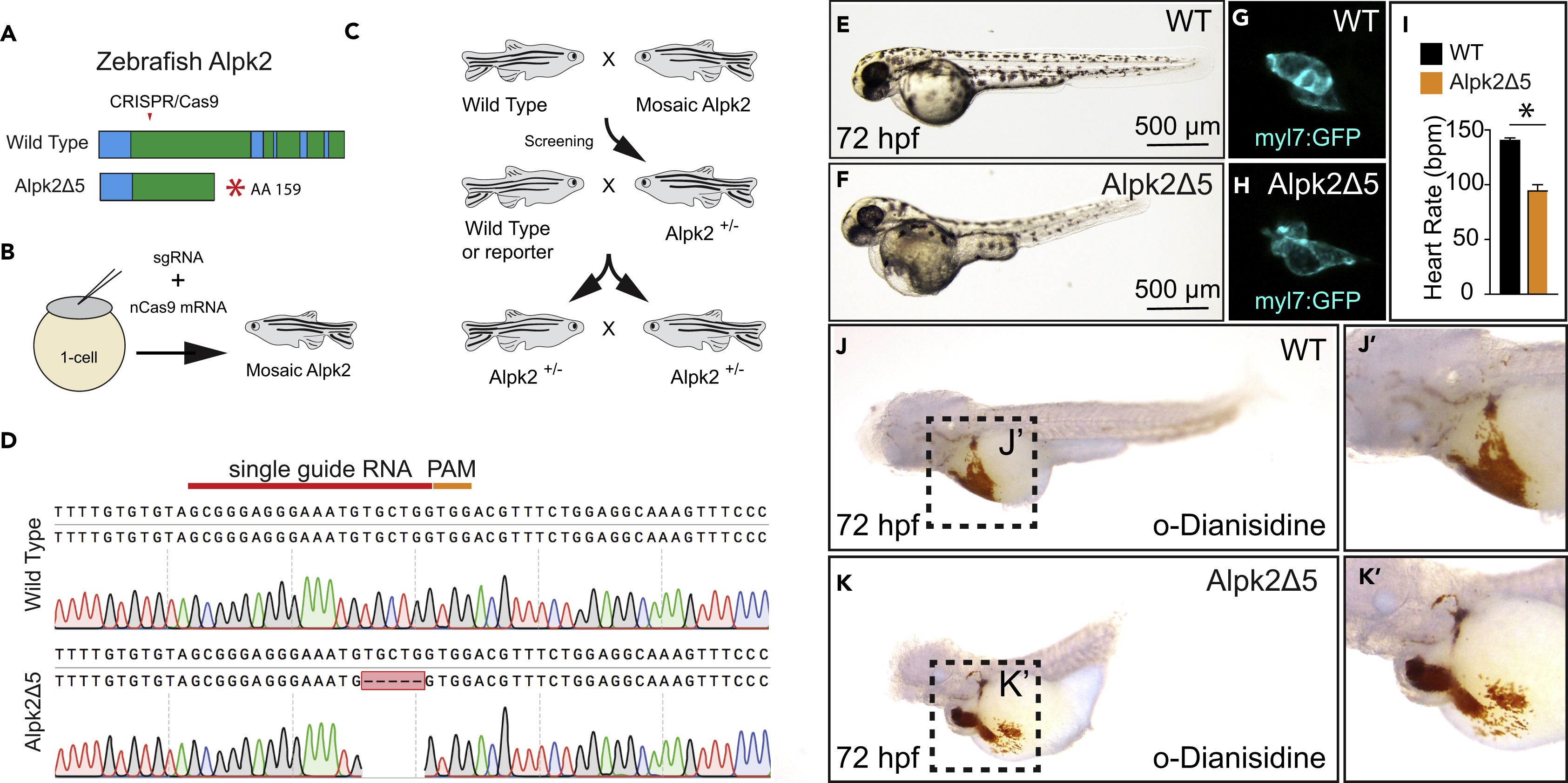
CRISPR/Cas9 Knockout of Zebrafish Alpk2 Inhibits Cardiac Development
(A) Schematic of the zebrafish Alpk2 locus and CRISPR/Cas9 targeted region. * denote stop codon.
(B and C) Injection and breeding paradigm to generate Alpk2 null homozygosity.
(D–H) (D) Sanger sequencing of homozygous mutation showing 5-bp deletion. Representative bright-field (E, F; 72 hpf) and fluorescent (G, H; 120 hpf) images of wild-type (E, G) and Alpk2 null zebrafish (F, H) carrying a transgene for myl7:GFP (G, H).
(I) Quantification of cardiac beating rate comparing Alpk2 null zebrafish to wild-type siblings at 72 hpf. * denotes p≤0.05.
(J and K) Representative bright-field images of hemoglobin-peroxidase staining with o-Dianisidine in 72 hpf WT and homozygous Alpk2Δ5 embryos. Data are representative of N ≥ 3 independent breeding experiments consisting of n∼30–150 embryos per spawn. hpf, hours post fertilization.

