
|
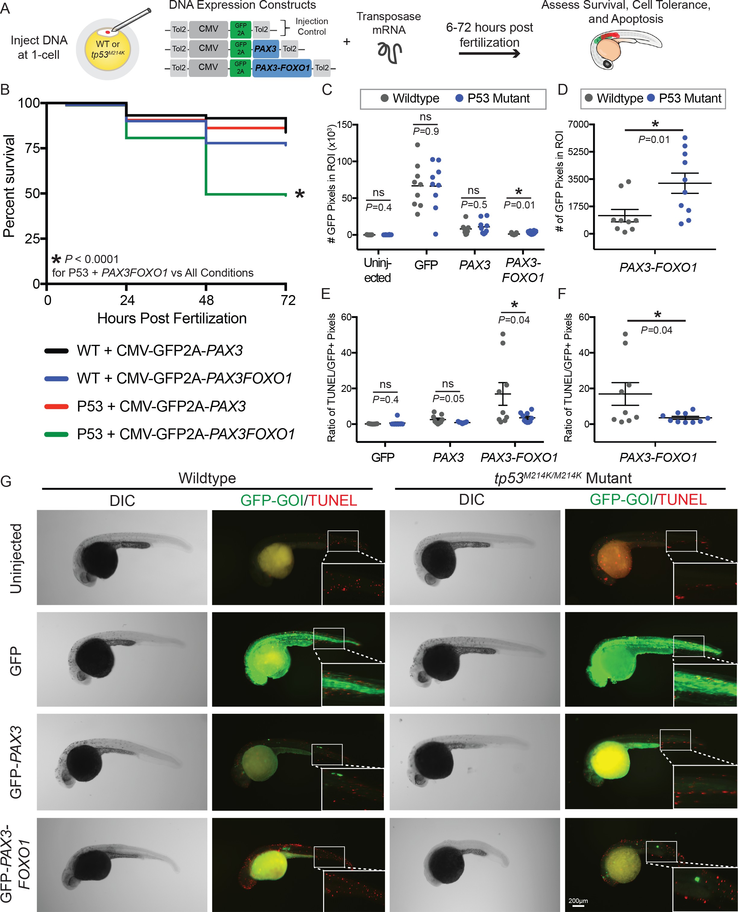
Fig. 2-S1
The tp53M214K mutation modifies the CMV-PAX3FOXO1 phenotype in developing zebrafish.
(A) Schematic of the experimental strategy to assess the impact of a tp53 mutation on survival, cell tolerance, and apoptosis of mosaic CMV restricted GFP, GFP-PAX3, or GFP-PAX3FOXO1 developmental expression. (B) Survival of PAX3 injected wildtype (n = 89) or tp53M214K/M214K (n = 146) as compared to PAX3-FOXO1 injected wildtype (n = 199) or tp53M214K/M214K (n = 219) evaluated at 6, 24, 48, and 72 hr post fertilization. All constructs were injected in equimolar amounts relative to 25 ng/µL of CMV-GFP2A-PAX3FOXO1. (C) Quantification of the number of GFP-positive pixels for each embryo imaged at 28 hr post fertilization using the same settings. Each marker represents a single zebrafish embryo, n = 8–10 embryos per group. Black bar is the mean, error bars represent SEM, and * indicates p<0.05, two-tailed Student’s t-test, ns- not significant. ROI- region of interest. (D) Same samples as in C plotted for the PAX3-FOXO1 injection groups only. (E) Quantification of TUNEL-positive pixels normalized to GFP-positive pixels indicated a lower proportion of PAX3-FOXO1 cells are undergoing apoptosis in the context of the tp53M214K/M214K mutation. Black bar is the mean, error bars represent SEM, n = 8–10 embryos per group, * indicates p<0.05, two-tailed Student’s t-test. (F) Same samples as in E plotted for the PAX3-FOXO1 injection groups only. (G) Representative images from wildtype and tp53M214K/M214K uninjected controls, CMV-GFP2A injection controls, CMV-GFP2A-PAX3 and CMV-GFP2A-PAX3FOXO1 injected experimental groups. Embryos were fixed at 28 hr post-injection, TUNEL (rhodamine) performed, and then embryos were counter-stained for GFP to denote transgene expression.