
Fig. 8
- ID
- ZDB-IMAGE-180807-8
- Publication
- Lai et al., 2018 - Liver-directed microRNA-7a depletion induces nonalcoholic fatty liver disease by stabilizing YY1-mediated lipogenic pathways in zebrafish
- All Figures
- Figures for Lai et al., 2018
|
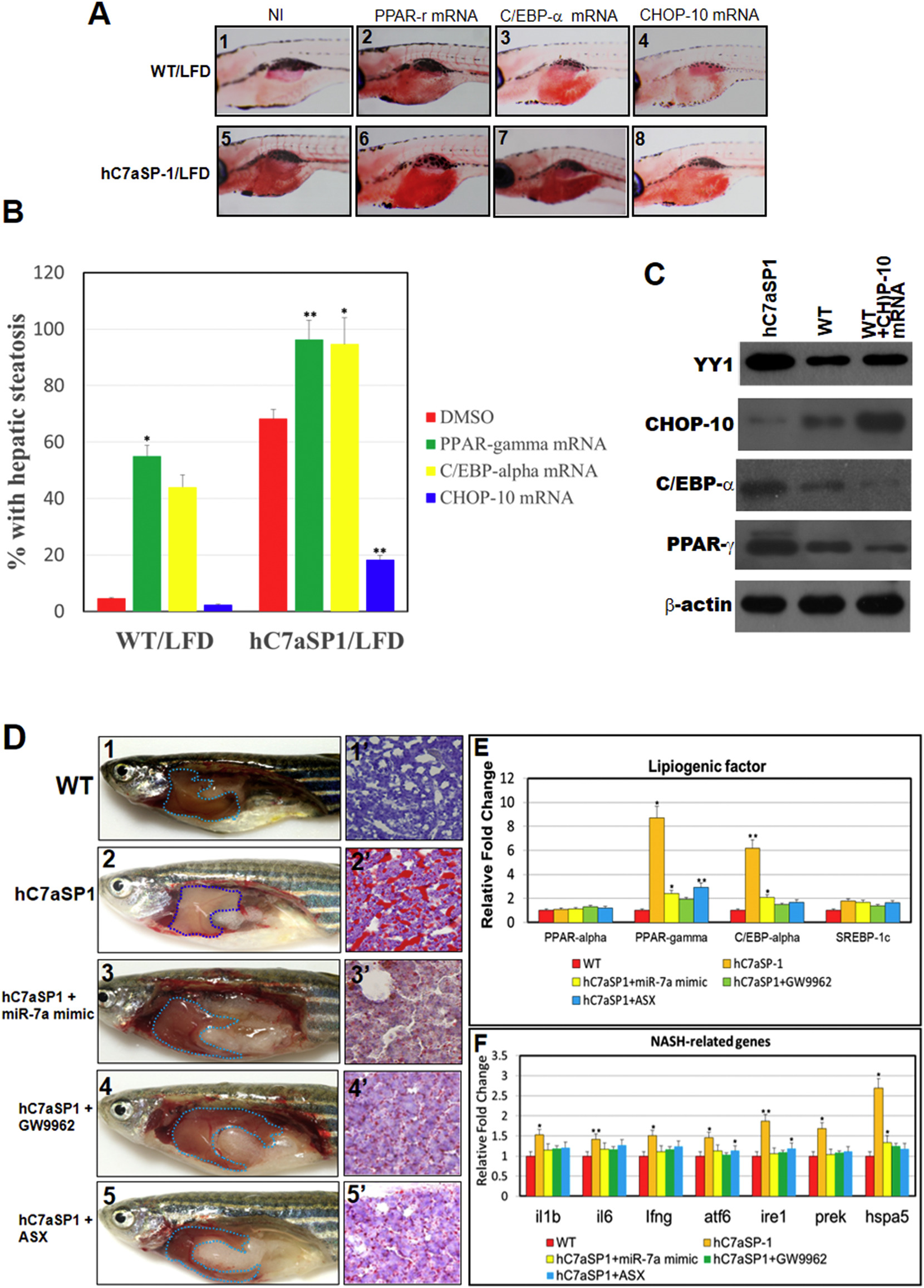
Fig. 8
PPAR-γ antagonists and miR-7a mimic treatment ameliorate NAFLD and NASH phenotypes in hC7aSP1 fed with LFD. (A) Steatotic analyses of 9 dpf WT and hC7aSP1r larvae. Embryos without injection (NI) and those injected with 200 pg PPAR-γ mRNA, 150 pg C/EBP-α mRNA, and 200 pg CHOP-10 mRNA were analyzed at the embryonic stage of 7 dpf. (B) Quantification of hepatic steatosis of WT and hC7aSP1 is shown in (A). The total number (N) of larvae in each group is indicated. (C) Western blot analyses of CHOP-10 ectopic efficiency. hC7aSP1 larvae and WT embryos without injection (NI) and those injected with 200 pg CHOP-10 mRNA were harvested at 9 dpf. β-Actin was used as the loading control. (D) Gross anatomy of liver and frozen liver section ORO staining in hC7aSP1 adults treated with miR-7a mimic, Gw9962, ASX, miR-7a mimic + GW9962, and miR-7a mimic + GW9962 (rescued hC7aSP1). (E) qRT-PCR analysis of selected lipogenic genes in the livers of rescued hC7aSP1. (F) qRT-PCR analysis of selected NASH-like genes in the livers of rescued hC7aSP1. Levels of mRNA were normalized to β-actin and expressed as fold of values in the WT control. **p < 0.01; *p < 0.05.