
Fig. S4
- ID
- ZDB-IMAGE-180730-39
- Publication
- Juan et al., 2018 - Myosin1D is an evolutionarily conserved regulator of animal left-right asymmetry
- All Figures
- Figures for Juan et al., 2018
|
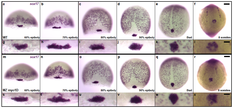
Fig. S4
Zebrafish myo1D is not required for the specification and migration of Left-Right organizer precursor cells a-x, sox17 in situ hybridization marks scattered endodermal cells as well as the posterior cluster of dorsal forerunner cells that give rise to the fish Left/Right Organizer, Kupffer’s Vesivle (KV). Comparison of WT (a-l) and MZ myo1D mutant (m-x) embryos reveals no differences in the number and behavior of KV precursor cells. a-d,m-p, are dorsal views, anterior up. e,f,q,r, are vegetal views of the tail bud region at the end of gastrulation (e,q) and the 8-somite stage (f,r). Anterior is up. a-f,m-r represent low magnification views of the whole embryo. g-l,s-x show high magnification views of the KV precursor cells. Scale bars: 50 μm in a-f,m-r . 20 μm in g-l,s-x.