
Fig. S6
- ID
- ZDB-IMAGE-180716-33
- Publication
- Kaur et al., 2018 - let-7 MicroRNA-Mediated Regulation of Shh Signaling and the Gene Regulatory Network Is Essential for Retina Regeneration
- All Figures
- Figures for Kaur et al., 2018
|
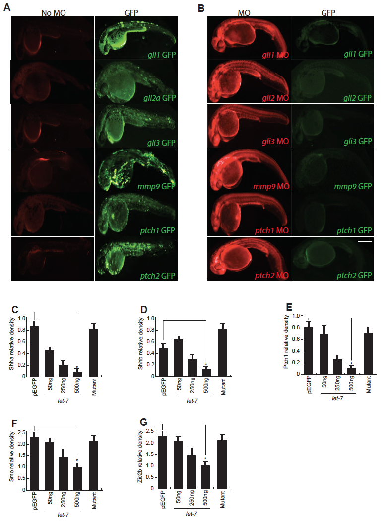
Fig. S6
MO assay in embryos. (A,B) The fusion mRNA, prepared by in vitro transcription using the clone containing GFP coding sequence in pCS2+ plasmid appended with the morpholino binding region of the respective genes, was injected alone (A), and along with morpholinos (B) in zebrafish embryos at single cell stage and imaged for GFP and lissamine fluorescence in a fluorescence microscope, at 24hpf. (C-G) Densitometry plots showing the expression of various GFP fusion proteins in let-7 micro RNA dependent manner in HEK293T cells, normalized to transfection control mCherry. *P<0.0001. Scale bars, 500 μm (A,B).