
IMAGE
Fig. 4
- ID
- ZDB-IMAGE-180712-60
- Genes
- Publication
- Kidwell et al., 2018 - Multiple zebrafish atoh1 genes specify a diversity of neuronal types in the zebrafish cerebellum
- All Figures
- Figures for Kidwell et al., 2018
Image
|
Figure Caption
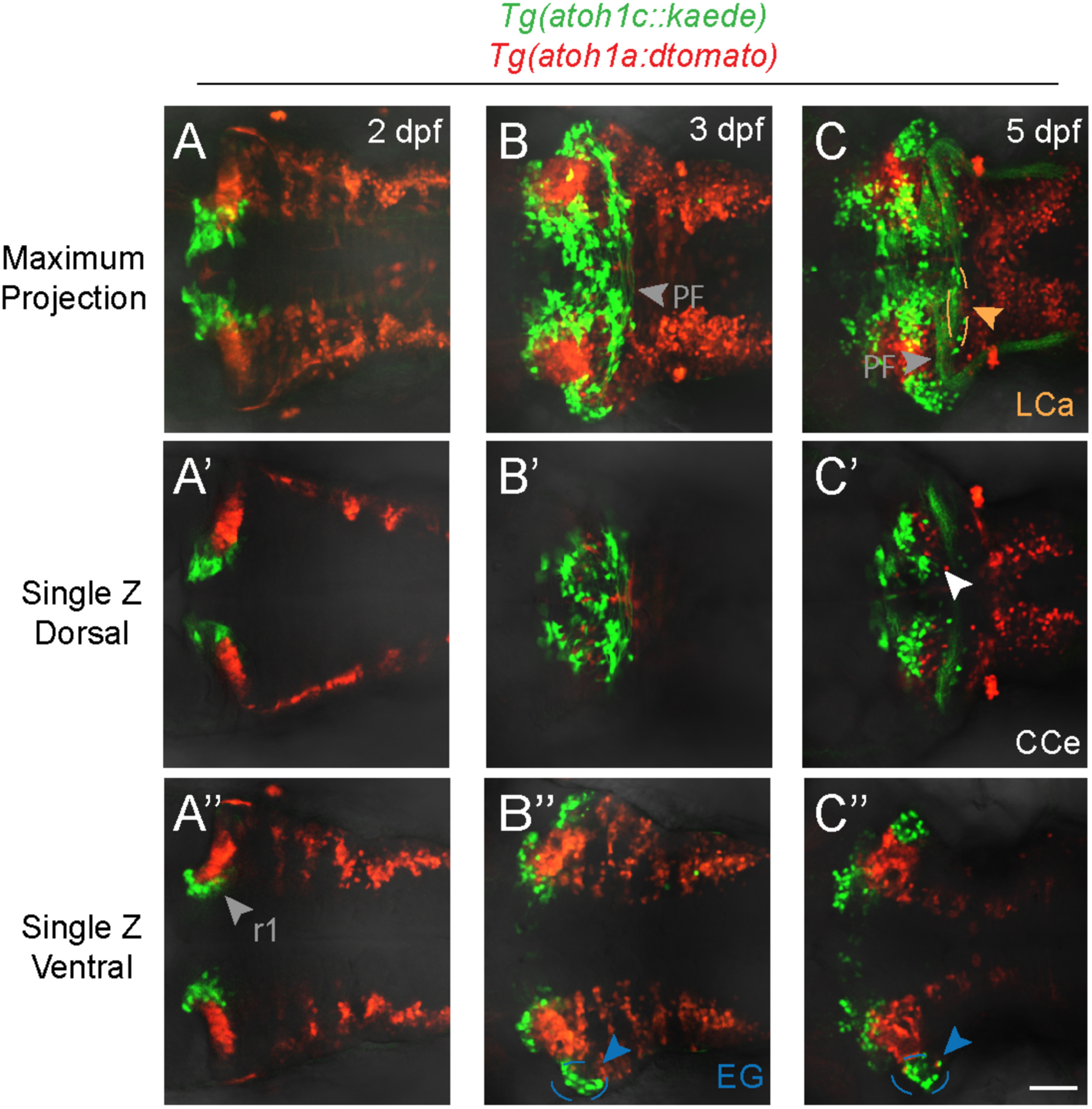
Fig. 4
atoh1a- and atoh1c-derived neurons specify distinct progenitor pools. Live imaging of wild-type embryos with Tg(atoh1c::kaede) (green) and Tg(atoh1a:dtomato) (red) transgenes indicate atoh1a and atoh1c-derived neurons are distinct cerebellar populations. Maximum projections of a z-stack (A-C) and single z-slices at dorsal (A′-C′) or ventral (A”-C”) focal planes with anterior to the left at time points as indicated. Scale bar: 50μM.
Figure Data
Acknowledgments
This image is the copyrighted work of the attributed author or publisher, and
ZFIN has permission only to display this image to its users.
Additional permissions should be obtained from the applicable author or publisher of the image.
Reprinted from Developmental Biology, 438(1), Kidwell, C.U., Su, C.Y., Hibi, M., Moens, C.B., Multiple zebrafish atoh1 genes specify a diversity of neuronal types in the zebrafish cerebellum, 44-56, Copyright (2018) with permission from Elsevier. Full text @ Dev. Biol.