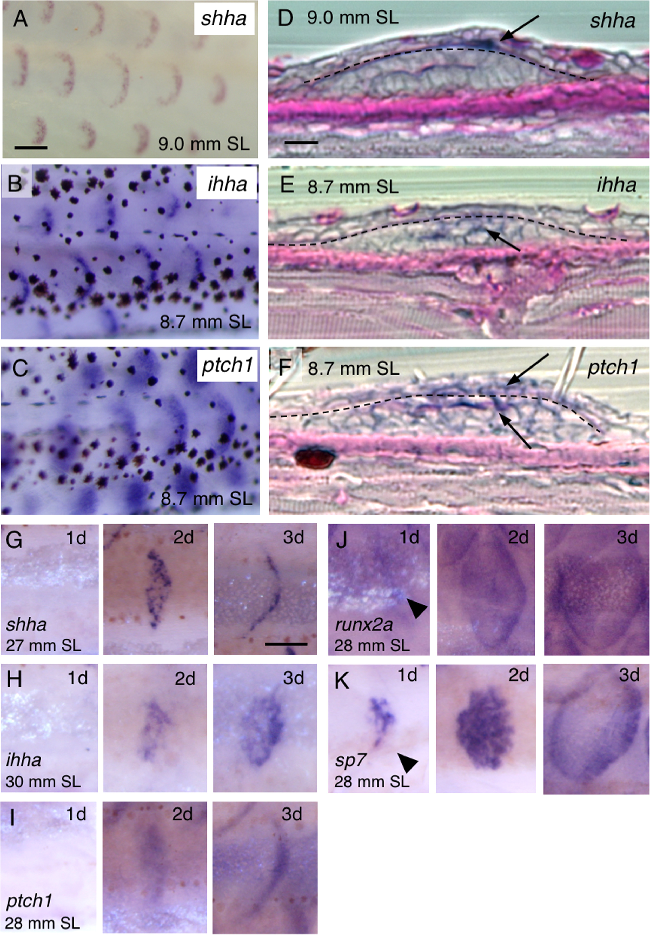
Fig. 5
Expression of hedgehog (Hh) signaling genes during development and regeneration. (A–C) Expression of sonic hedgehog a (shha) (A), indian hedgehog homolog a (ihha) (B), and patched 1 (ptch1) (C) in repetitive crescent-shaped domains along the trunk of juvenile fish; each domain presumably corresponds to a scale. mitfa–/– fish (nacre, devoid of pigmentation) were used in (A). (D–F) Histological sections of juvenile fish labeled with shha (D), ihha (E), and ptch1 (F) together with periodic acid-Schiff (PAS) staining. shha is predominantly expressed in epidermal cells (arrow in D), whereas ihha is primarily detected in the dermis (arrow in E), and ptch1 is broadly expressed in both the epidermis and dermis (arrows in F). Dashed lines indicate the basement membranes. (G–K) Expression of Hh signaling genes and osteoblast marker genes during regeneration. Scales were removed from the trunk of adult fish and then stained with anti-sense RNA probes for shha (G), ihha (H), ptch1 (I), runx2a (J), and sp7 (K) on Days 1–3 after scale removal. Expression of Hh signaling genes starts within 2 days (G–I), whereas, runx2a and sp7 are already expressed after 1 day (arrowheads in J,K). Scale bars, 100 µm (A–C), 10 µm (D–F), 200 µm (G–K).
Reprinted from Developmental Biology, 437(2), Iwasaki, M., Kuroda, J., Kawakami, K., Wada, H., Epidermal regulation of bone morphogenesis through the development and regeneration of osteoblasts in the zebrafish scale, 105-119, Copyright (2018) with permission from Elsevier. Full text @ Dev. Biol.

