
|
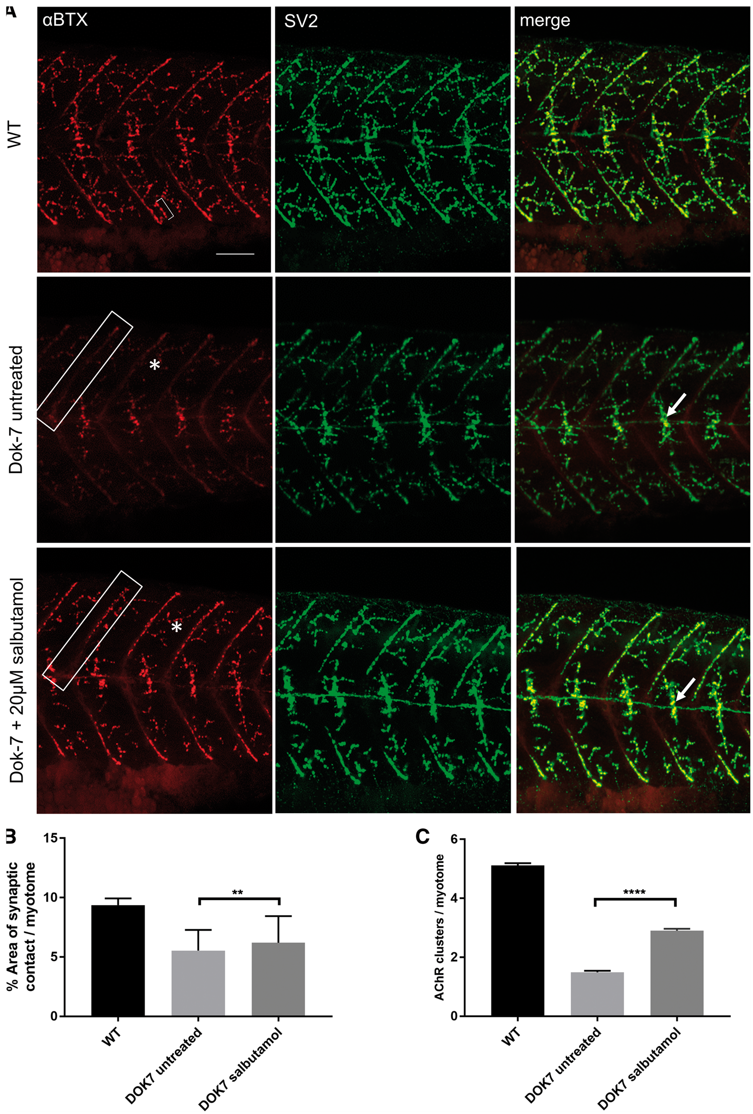
Fig. 2
Salbutamol treatment of Dok-7 MO-injected zebrafish improves NMJ morphology. Lateral views of 48 hpf embryos with neuromuscular synapses labelled with antibodies against SV2 (green, presynaptic vesicles) and αBTX (red, postsynaptic AChRs). Scale bar = 50 µm. (A) In Dok7 MO-injected embryos, the focal innervation point along the horizontal myoseptum is reduced in size compared with wild type, but is increased following treatment with 20 µm salbutamol (arrows). In addition, the morphology of myoseptal (boxes) and myotomal (asterisks) AChR clusters is partially rescued with salbutamol treatment. A representative 20 μm2 AChR cluster is labelled with a bracket. (B) Following salbutamol treatment, the area of synaptic contact on the horizontal myoseptum is significantly increased (** indicates P < 0.01, Student’s t-test, n = 160 treated and 160 untreated myotomal segments examined). (C) Salbutamol treatment caused a significant increase in the number of AChR clusters >20 μm2 per myotome in Dok7 zebrafish embryos (**** indicates P < 0.0001, Student’s t-test, n = 100 treated and 100 untreated myotomal segments examined).