
Image
|
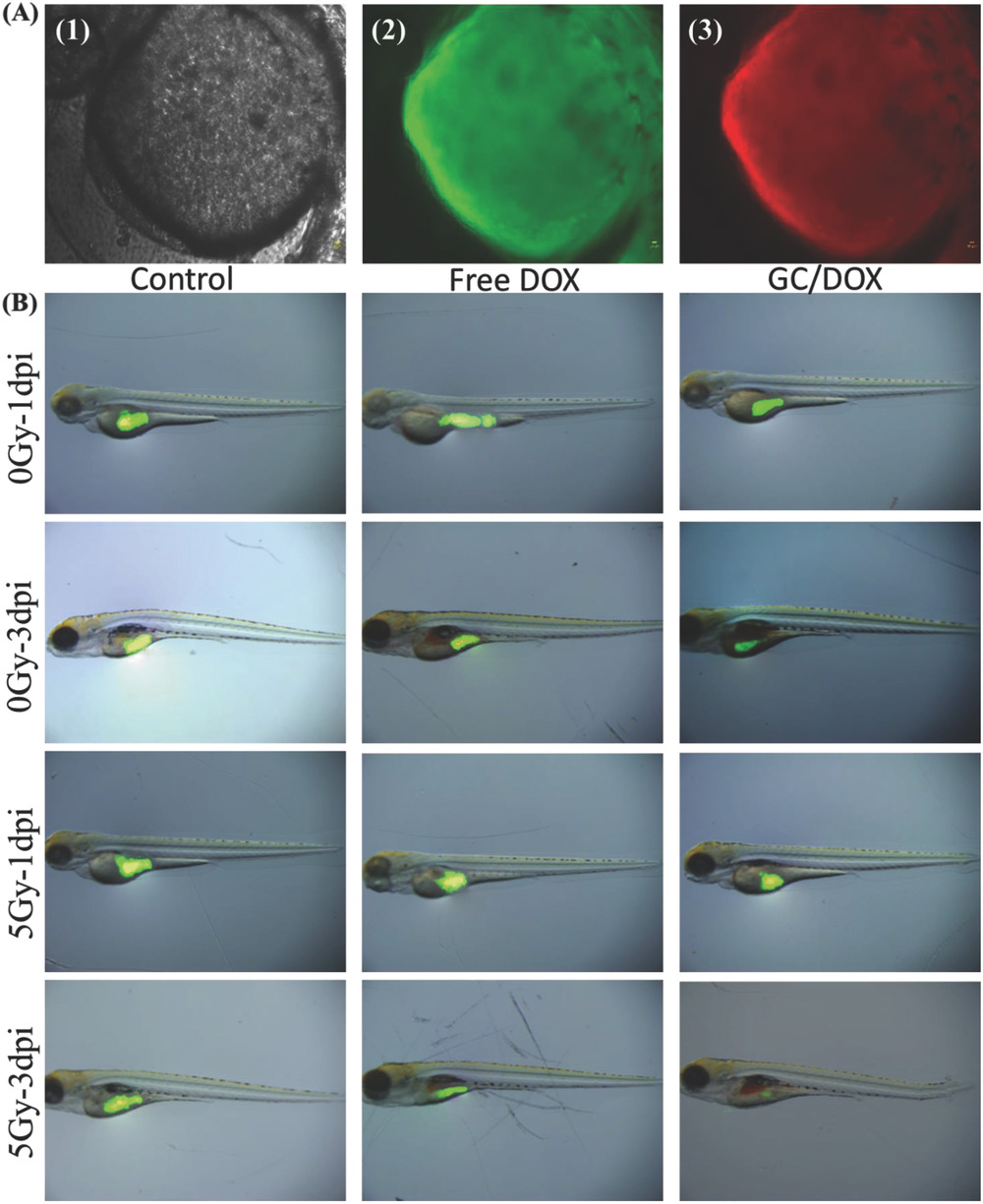
Figure Caption
Fig. 8
In vivo zebrafish study. A) 2‐dpf zebrafish were exposed to GC/DOX for 24 h; bright‐field microscopy (1), green fluorescence (2), and red fluorescence (3). B) HeLa cells were labeled with CFSE and treated with free DOX and GC/DOX for 48 h. CFSE fluorescence was recorded 0 and 3 dpi (in the presence and absence of γ‐radiation).
Acknowledgments
This image is the copyrighted work of the attributed author or publisher, and
ZFIN has permission only to display this image to its users.
Additional permissions should be obtained from the applicable author or publisher of the image.
Full text @ Adv Sci (Weinh)