
|
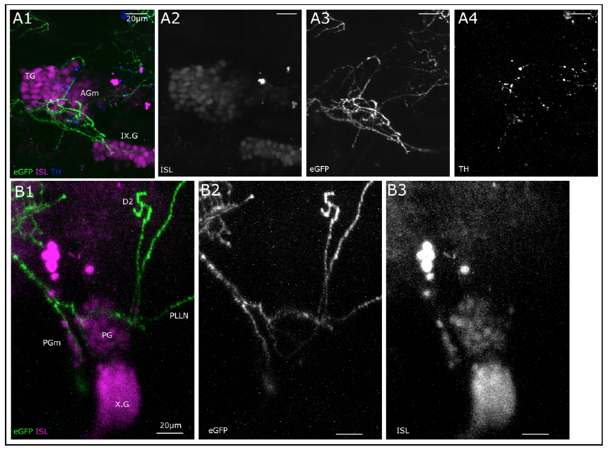
Fig. S1
Dopaminergic innervation of lateral line ganglia.
A. Lateral view, MIP (total depth of 54.19 μm, step size: 1.18 μm), of region of medial anterior lateral line ganglion (mAG) with trigeminal ganglion (TG) and glossopharyngeal ganglion (IX.G). GFP expression driven by th:Gal4-VP16 (green, A3) in projections targeting the trigeminal and lateral line ganglion and also running through IX.G. Cell bodies of sensory afferent neurons in ganglia marked with Islet1/Islet2 immunoreactivity (ISL, magenta, A2). TH-immunoreactivity shown in blue and A4. B. Lateral view, MIP (total depth of 40.95 μm, step size 1.17 μm), of region of posterior lateral line ganglion, which can be further subdivided into medial posterior lateral line ganglion (PGm) and posterior lateral line ganglion (PG). Also visible: caudal vagus ganglion (X.G). Neurons with GFP expression (green and B2) target PGm and PG and project to dorsal neuromast, along central projection of lateral line afferent neurons and posterior lateral line nerve (PLLN). Sensory afferent neurons marked with ISL-immunoreactivity (magenta and B3). All scale bars: 20μm.