
Fig. 2
- ID
- ZDB-IMAGE-180703-11
- Publication
- Myllymäki et al., 2018 - Identification of protective postexposure mycobacterial vaccine antigens using an immunosuppression-based reactivation model in the zebrafish
- All Figures
- Figures for Myllymäki et al., 2018
|
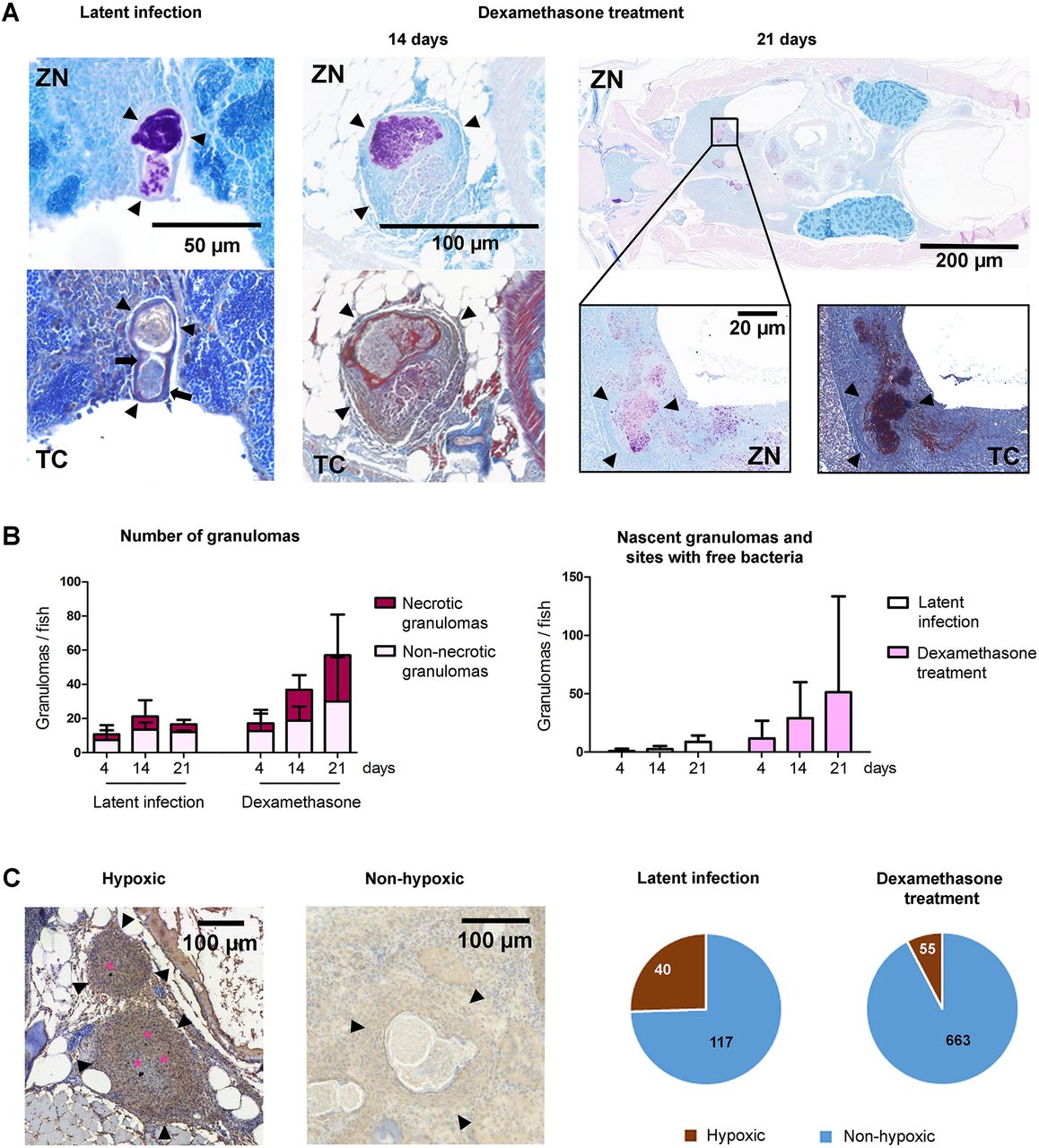
Fig. 2
Dexamethasone treatment leads to disruption of the granuloma structure, increase in the number of granulomas and loss of hypoxia in zebrafish with a latent mycobacterial infection. (A) Ziehl–Neelsen staining (ZN) of mycobacteria (purple). Black arrowheads indicate the outline of a granuloma. Trichrome staining (TC) of fibrous tissue around granulomas (blue) (black arrows). (B) Quantification of the total number of granulomas and the proportion of necrotic and non-necrotic granulomas (left), and the number of nascent granulomas and sites with non-capsulated mycobacteria (right) per fish after 4, 14 and 21 days of dexamethasone treatment or normal feeding (n=3-6 fish/group). Data are presented as mean±s.d. Statistical significance was assessed by unpaired Student's t-test. (C) Hypoxic staining shows hypoxic lesions inside granulomas in dark brown (pink stars). The proportions of hypoxic and non-hypoxic granulomas during a latent infection, and after 2 weeks of dexamethasone treatment, are quantified in the pie chart. See also Fig. S2.